Advertisement

Displaying posts in the category: News

New partnership with MicroscopyDB

Posted by , on 30 October 2023

We are delighted to announce our new partnership with MicroscopyDB – a global repository of online microscopy resources. This new partnership means that the listings on our job and events boards will be pulled in from the MicroscopyDB database, ensuring that FocalPlane readers can keep up to date with event and job listings across the

Featured image with Ioakeim (Makis) Ampartzidis

Posted by , on 27 October 2023

Our featured image, ‘Dandelion in spring’ from Ioakeim (Makis) Ampartzidis, shows neuroepithelial-like cells cultured on glass beads. The cell nuclei are coloured in magenta, and the cytoskeleton is stained in black, labelling F-actin. The actively proliferating cells are highlighted with Ki-67 antigen in green. We caught up with Makis to find out more about his

Featured image with Victoria Alonso

Posted by , on 13 October 2023

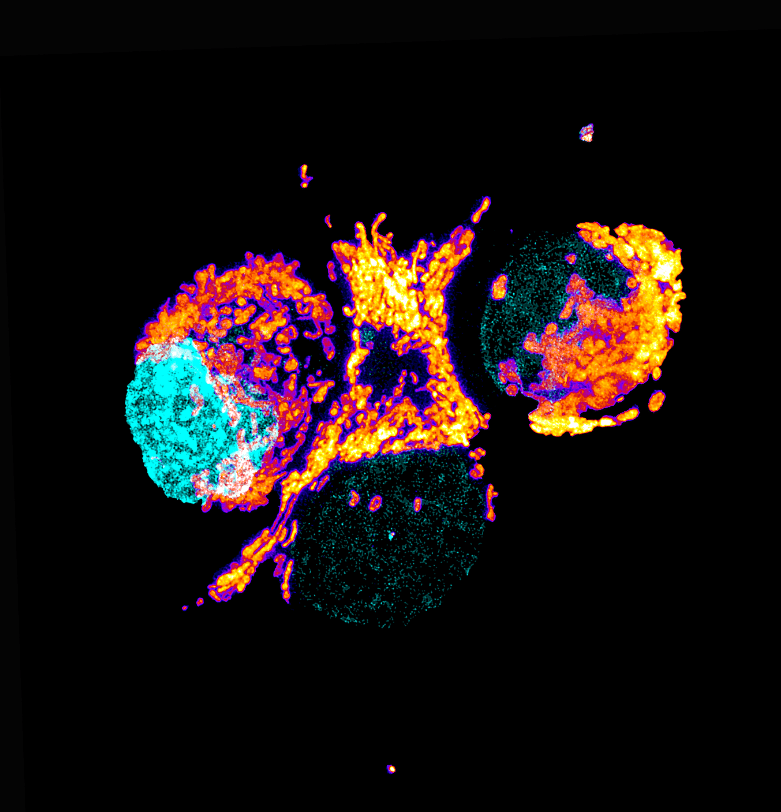
Our featured image is a Trypanosoma cruzi epimastigote, prepared for expansion confocal microscopy with pan-proteome staining with NHS-ester Atto564 by Victoria Alonso. The image is a z-stack acquired with a Zeiss LSM 880 and was edited with ImageJ Fiji, plugin: Z-stack depth Colorcode.

Becoming ‘Greener’ with Light Microscopy

Sponsored by CoolLED, on 11 October 2023

Working in a laboratory, you can’t help but notice how everyday tasks can impact the environment – from reliance on single-use plastics to energy-hungry equipment. While this might seem unavoidable, sustainable options are becoming widely available. In fact, many technologies and initiatives now exist to support the scientific community on the journey towards sustainable laboratories.  

Featured image with Peggy Paschke

Posted by , on 29 September 2023

Our featured image is called Dictyostelium Trees on Fire and was acquired by Peggy Paschke. The image shows Dictyostelium discoideum cells stably expressing the cAMP sensor Flamindo2. Cells were imaged as 5×5 tile scan for 5 hours with a Nikon AXR (488 nm excitation) using a 4x air objective. Shown is a cropped part of

The FocalPlane Network: joining and using the database

Posted by , on 20 September 2023

Our instructional videos take you step-by-step through the process of joining and using the FocalPlane Network. You can contact us at focalplane@biologists.com with any feedback on the Network. Joining the FocalPlane Network Using the FocalPlane Network

The FocalPlane Network: connecting the microscopy community

Posted by , on 20 September 2023

The FocalPlane Network is an international directory of researchers with expertise in microscopy. The goal of the Network is to facilitate promotion and networking, as well as assisting those seeking conference speakers, committee members, reviewers or collaborators. Membership is open to researchers in the microscopy community from all career stages, geographical locations or areas of

Featured image with Martín Estermann

Posted by , on 15 September 2023

Our featured image, acquired by Martín Estermann, is a 3D reconstruction of C. elegans transgenic embryos. Embryos were imaged live using a Zeiss Celldiscoverer 7 microscope. The 3D reconstruction was performed using Fiji. Histones in are shown in green and the cell membrane in magenta. We caught up with Martín to find out about his

And the winner is...

Posted by , on 1 September 2023

We are delighted to announce that the winner of the 2023 FocalPlane image competition is Rebecca Simkin. Rebecca’s image, ‘Neuromuscular junctions’, depicts four NMJs in a lumbrical muscle, located in the hind paw of a wildtype mouse. A wholemount immunostaining was performed using 2H3 and SV2 antibodies to visualise lower motor neurons in green (specifically

Unveiling the Marvels of the First-Ever Gordon Research Conference on Volume Electron Microscopy - Capturing Subcellular Architecture from Organelles to Organisms -By Ana Correia and James Thompson

Posted by , on 24 August 2023

We delve into our captivating journey at the first Gordon Research Conference (GRC) on Volume Electron Microscopy (vEM) as early career scientists. Set against the backdrop of sunny Ventura, California, this conference brought together experts in the vEM field from all corners of the globe, creating an intellectual haven for learning, networking, and forging collaborations.