Advertisement

Your search returned 10 posts.

Featured image with Joe McKellar

Posted by , on 8 December 2023

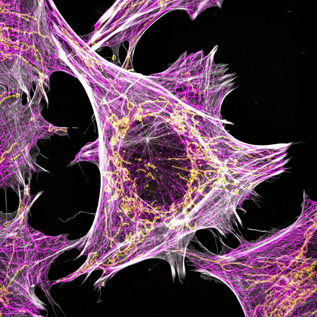
Our featured image shows a culture of boa constrictor kidney cells that have been transfected with a fusogenic viral protein. This leads to the formation of giant multi-nucleated cells. The cells were transfected with the fusogenic viral protein (shown in gold), left several days for efficient protein expression and for cell fusion to occur, plated

Featured image with Nick Gatford

Posted by , on 16 November 2023

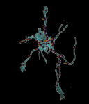
Our featured image shows a single human dopaminergic neuron generated from a human stem cell, acquired via super-resolution Airyscan confocal microscopy at the University of Oxford Micron facility by Nick Gatford. We caught up with Nick to find out about his research and what he is excited about in microscopy.

Featured image with Oona Paavolainen

Posted by , on 15 November 2023

Our featured image, acquired by Oona Paavolainen, shows a primary mammary gland organoid, in a collagen-I matrix, formed from a single primary epithelial cell isolated from material obtained from a breast reduction surgery. The image was taken with a spinning disk confocal microscope (3i CSU-W1 Spinning disk) using a 20x Zeiss Plan-Apochromat (NA=0.8) objective. The

Featured image with Till Stephan

Posted by , on 14 November 2023

Our featured image, acquired by Till Stephan, shows a live-cell stimulated emission depletion (STED) microscopy recording of a HeLa cell in which mitochondria (orange-magenta) and microtubules (cyan) are labelled. The super-resolution image reveals the densely stacked mitochondrial cristae, coordinated invaginations of the mitochondrial inner membrane.

Featured image with Ioakeim (Makis) Ampartzidis

Posted by , on 27 October 2023

Our featured image, ‘Dandelion in spring’ from Ioakeim (Makis) Ampartzidis, shows neuroepithelial-like cells cultured on glass beads. The cell nuclei are coloured in magenta, and the cytoskeleton is stained in black, labelling F-actin. The actively proliferating cells are highlighted with Ki-67 antigen in green. We caught up with Makis to find out more about his

Featured image with Victoria Alonso

Posted by , on 13 October 2023

Our featured image is a Trypanosoma cruzi epimastigote, prepared for expansion confocal microscopy with pan-proteome staining with NHS-ester Atto564 by Victoria Alonso. The image is a z-stack acquired with a Zeiss LSM 880 and was edited with ImageJ Fiji, plugin: Z-stack depth Colorcode.

Featured image with Peggy Paschke

Posted by , on 29 September 2023

Our featured image is called Dictyostelium Trees on Fire and was acquired by Peggy Paschke. The image shows Dictyostelium discoideum cells stably expressing the cAMP sensor Flamindo2. Cells were imaged as 5×5 tile scan for 5 hours with a Nikon AXR (488 nm excitation) using a 4x air objective. Shown is a cropped part of

Featured image with Martín Estermann

Posted by , on 15 September 2023

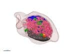
Our featured image, acquired by Martín Estermann, is a 3D reconstruction of C. elegans transgenic embryos. Embryos were imaged live using a Zeiss Celldiscoverer 7 microscope. The 3D reconstruction was performed using Fiji. Histones in are shown in green and the cell membrane in magenta. We caught up with Martín to find out about his

Featured image with Beste Senem Değirmenci

Posted by , on 18 August 2023

Our featured image, ‘Bubbles of The Cell’, shows enlarged endosomal vesicles in HeLa S3 cells upon inhibition of PI(3,5)P2 activity. PI(3,5)P2 is a crucial phosphoinositide involved in the regulation of endosomal trafficking. Depletion of PI(3,5)P2 disrupts the transition from early to late endosomes, leading to the enlargement of endosomal vesicles. Imaged with Leica DMI8 SP8

Featured image with Outi Paloheimo

Posted by , on 4 August 2023

Our featured image is laser scanning confocal microscope image showing cell division of Madin-Darby canine kidney cells with nuclei in grey and filamentous actin in orange. The image was acquired with a Zeiss LSM 780 microscope with a 63x/1.40 oil immersion objective, 405 nm diode laser and 561 nm diode-pumped solid-state laser. Deconvolution was performed