Featured image with Ellen C. O’Shaughnessy
Posted by FocalPlane, on 31 January 2025
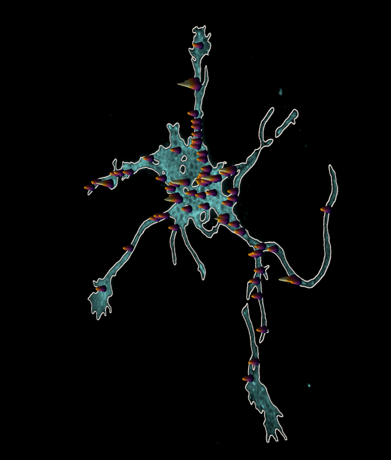
Our featured image shows the locations of exocytic hotspots in a developing cortical neuron. Ellen O’Shaughnessy, Stephanie Gupton and colleagues developed pHusion, a toolkit for analysis of exocytosis that was published in Journal of Cell Science’s special issue on ‘Imaging Cell Architecture and Dynamics‘. You can learn more about pHusion in this tutorial from the Gupton lab.
Find out more about Ellen’s research below:
Research career so far: I began my career in synthetic biology studying the emergent properties of signalling pathways and how tuning component properties such as expression level or localization can give rise to very different systems-level behaviours. My interest in signalling led to further work developing and utilizing biosensors of small GTPase activity in various live cell systems. In this work, I was able to explore both 2D and 3D imaging modalities and dive deeply into image analysis and computer vision.
Current research: I am currently studying membrane remodelling in developmental neurons and building computational tools for the unbiased detection of exocytic events, analysis of the spatio-temporal dynamics of these events and characterization of membrane movement during morphogenesis.
Favourite imaging technique/microscope: I have most enjoyed using lattice-light sheet microscopy (LLSM) for the incredible balance of speed and resolution with minimal cell perturbation. I used this technique to measure the activity of a specific GTPase in rapidly moving cells and to capture the phenomenal transitions megakaryocytes (MKs) undergo in generating platelets. During proplatelet formation, MKs transform from very large round cells into a vast array of long, branched, and highly dynamic extensions which are beautiful to watch in real time.
What are you most excited about in microscopy? I am most excited by the development of numerous microscopy techniques that break the diffraction limit and, particularly, how these can be implemented in live cells. I am predominantly interested in the dynamics of cell processes and how live cells respond to their environments, which is very difficult to study in fixed samples. Techniques such as Airyscan, SIM, STED and others are being used currently to achieve higher resolution in live cells and I am excited to see how much better these systems will become over time.
 (No Ratings Yet)