Advertisement

Your search returned 10 posts.

Featured image with Jovan Brockett

Posted by , on 19 July 2024

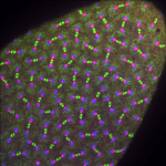
Our featured image, acquired by Jovan Brockett, shows a Drosophila melanogaster embryo undergoing syncytial nuclear divisions during early embryonic development. It was prepared using PFA fixation and hand dissected to remove the outer vitelline membrane. The image was captured using a Nikon Ti-E system fitted with a Yokagawa CSU-X1 spinning disk head, Hamamatsu Orca Flash

Featured image with Hoang Anh Le

Posted by , on 5 July 2024

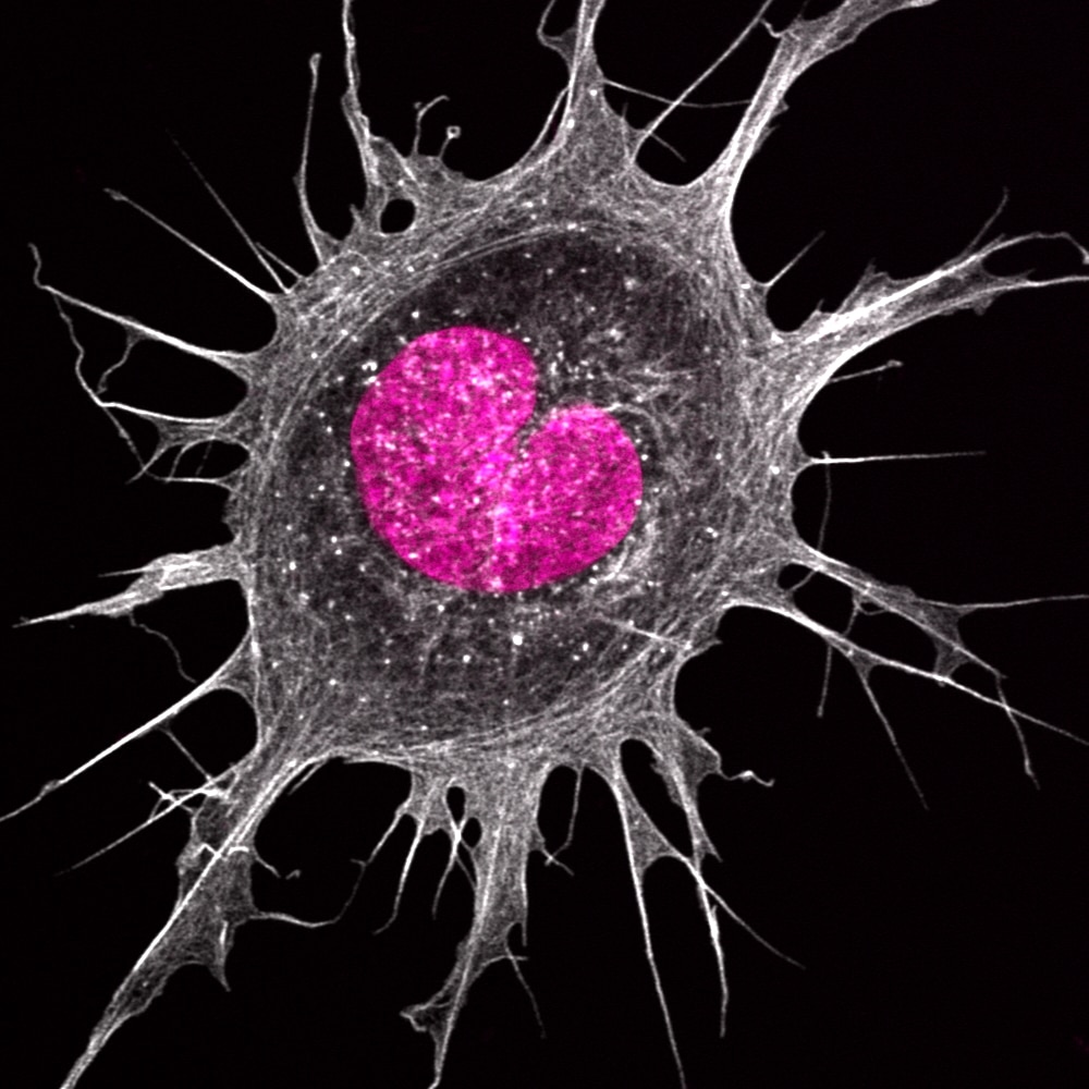
Our featured image, acquired by Hoang Anh Le, shows an Ewing’s sarcoma cancer cell plated on a fibronectin-coated surface. The cell was fixed with 4% formaldehyde and stained using phalloidin (white) and DAPI (in pink). The sample was imaged using a confocal Zeiss 880 microscope with Airyscan.

Featured image with Theresa Wiesner

Posted by , on 21 June 2024

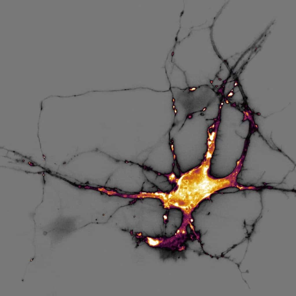
Our featured image, acquired by Theresa Wiesner, shows a neuron in dissociated hippocampal culture and was imaged using highly inclined thin illumination microscopy (HiLO). The neuron is transfected with a neurotransmitter (glutamate) sensor and shown in orange. The image was acquired using Nikon Elements software / microscope and post-processing was done using FIJI (ImageJ). Find

Featured image with Jen Silverman

Posted by , on 24 May 2024

Our featured image, acquired by Jen Silverman, shows a lateral view of a tuft cell taken from a frozen tissue section and imaged using structured illumination microscopy (SIM). F-actin, labelled using phalloidin, is shown in cyan and an actin bundling protein enriched in tuft cells is shown in orange. Reconstruction of the image was performed

Featured image with Sujan Ghimire

Posted by , on 26 April 2024

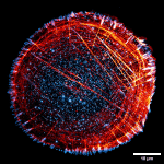
Our featured image, acquired by Sujan Ghimire, shows a U2OS cell stained for paxillin (Cyan Hot) and actin (Red Hot) following an immunofluorescence staining protocol. The image was captured using structured illumination microscopy (SIM) with Deltavision OMX (63x Oil, NA: 1.42) at Cell Imaging and Cytometry facility, Turku Bioscience.

Featured image with Thibault Dhellemmes and Jérémie Teillon

Posted by , on 1 March 2024

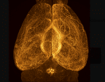
Our featured image shows the full morphology of Relaxin-3 neurons in an entire adult mouse brain. To observe the full depth of this brain, it was made transparent by applying an optical clearing technique called Adipoclear. The sample was observed on an Ultramicroscope II light sheet microscope and is a maximum intensity projection over the

Featured image with Rossana Melo

Posted by , on 16 February 2024

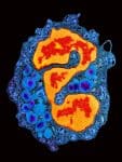
Our featured image, acquired by Rossana Melo, shows a human eosinophil isolated from the peripheral blood using a negative immunomagnetic selection method and prepared for transmission electron microscopy (TEM). The image was acquired with a Tecnai G2 Spirit Bio Twin ThermoFisher Scientific (former FEI) electron microscope.

Featured image with Virginia Barrera

Posted by , on 2 February 2024

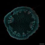
Our featured image was acquired by Virginia Barrera and was the winning image from the Institute of Molecular and Cellular Biology of Rosario (IBR-CONICET) annual retreat. It depicts a transversal cut of a soybean, with Calcofluor White staining cellulose and Basic Fuchsine labelling lignin.

Featured images in 2023

Posted by , on 5 January 2024

In 2023, we started a featured image series and we have been delighted to highlight the microscopists that acquired these beautiful images! You can check out the full series here and in the gallery below. We are continuing the series in 2024 and would love to have nominations, including self-nominations. Send us an email at

Featured image with Julien Rességuier

Posted by , on 22 December 2023

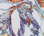
Our featured image, acquired by Julien Rességuier, shows the branchial cavity lymphoid architecture of an adult zebrafish. It is a 2D projection of a 3D multi-field of view acquisition of a transversal cryosection (30 μm) that has been stained with fluorescent phalloidin (inverted LUT – grey/copper) and DAPI (blue) to reveal tissue structures and labelled