Advertisement

Featured image with Ioakeim (Makis) Ampartzidis

Posted by , on 27 October 2023

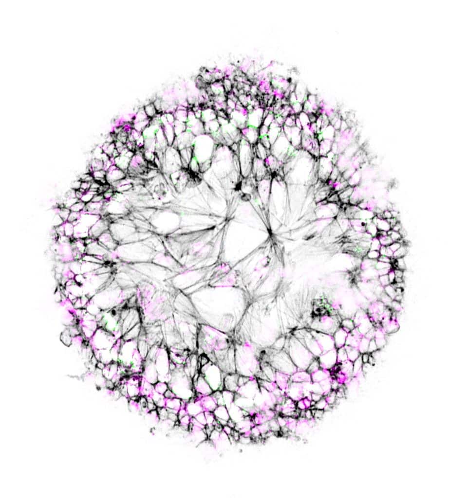
Our featured image, ‘Dandelion in spring’ from Ioakeim (Makis) Ampartzidis, shows neuroepithelial-like cells cultured on glass beads. The cell nuclei are coloured in magenta, and the cytoskeleton is stained in black, labelling F-actin. The actively proliferating cells are highlighted with Ki-67 antigen in green. We caught up with Makis to find out more about his

Microscopy preprints – new tools and techniques in imaging

Posted by , on 20 October 2023

Here is a curated selection of preprints posted recently on new tools and techniques in imaging. Let us know if we are missing any preprints that are on your reading list.

Become a FocalPlane correspondent

Posted by , on 17 October 2023

Enthusiastic about science communication and looking for a chance to broaden your writing experience alongside your research activities? FocalPlane, our community site for microscopists, is looking to appoint three scientists as correspondents who will play a key role in developing and writing content over the coming year. In 2023, we have been working with Mariana De

Euro-BioImaging has a Scientific Ambassador program!

Posted by , on 16 October 2023

We are delighted to announce that Euro-BioImaging has launched a Scientific Ambassador program. This program will raise awareness about the services and opportunities provided by Euro-BioImaging, by engaging with new communities.

Featured image with Victoria Alonso

Posted by , on 13 October 2023

Our featured image is a Trypanosoma cruzi epimastigote, prepared for expansion confocal microscopy with pan-proteome staining with NHS-ester Atto564 by Victoria Alonso. The image is a z-stack acquired with a Zeiss LSM 880 and was edited with ImageJ Fiji, plugin: Z-stack depth Colorcode.

Becoming ‘Greener’ with Light Microscopy

Sponsored by CoolLED, on 11 October 2023

Working in a laboratory, you can’t help but notice how everyday tasks can impact the environment – from reliance on single-use plastics to energy-hungry equipment. While this might seem unavoidable, sustainable options are becoming widely available. In fact, many technologies and initiatives now exist to support the scientific community on the journey towards sustainable laboratories.  

Microscopy preprints – bioimage analysis tools

Posted by , on 6 October 2023

Here is a curated selection of preprints published recently. In this post, we focus specifically on new tools for bioimage analysis.

behind DeXtrusion: an automated pipeline to detect epithelial cell extrusion through machine learning

Posted by , on 6 October 2023

The problem In animals, epithelial tissue covers most of our organs. In order to create the very intricate and complex shapes of these organs, these tissues are drastically remodelled during embryogenesis through growth and shape changes. Once they achieve these final shapes, they maintain their cell number in a dynamic equilibrium. Both of these mechanisms,

Featured image with Peggy Paschke

Posted by , on 29 September 2023

Our featured image is called Dictyostelium Trees on Fire and was acquired by Peggy Paschke. The image shows Dictyostelium discoideum cells stably expressing the cAMP sensor Flamindo2. Cells were imaged as 5×5 tile scan for 5 hours with a Nikon AXR (488 nm excitation) using a 4x air objective. Shown is a cropped part of

How to… Publish Images blog series

Posted by , on 27 September 2023

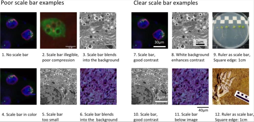
by Helena Jambor In biology, images document appearances or structures of animals, issues, or cells, and increasingly are a source of quantitative insights of biological processes. Many papers are published every single day with images (Lee, 2018).Not all of these images are truthful or insightful. Some images are manipulated to mislead audiences (Bik et al.,