Featured image with Peggy Paschke
Posted by FocalPlane, on 29 September 2023
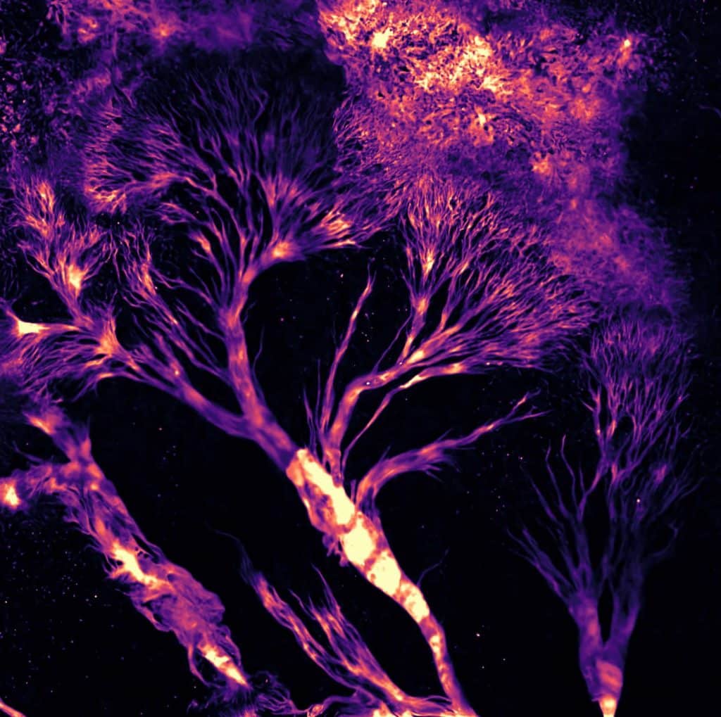
Our featured image is called Dictyostelium Trees on Fire and was acquired by Peggy Paschke. The image shows Dictyostelium discoideum cells stably expressing the cAMP sensor Flamindo2. Cells were imaged as 5×5 tile scan for 5 hours with a Nikon AXR (488 nm excitation) using a 4x air objective. Shown is a cropped part of the final tile scan, at the very edge of the agar slope, which creates the elution of Dicty Trees. Tile scans were stitched together using ImageJ. LUT is mpl magma.
We caught up with Peggy to find out about her research and what she is excited about in microscopy.
Research career so far: I did my PhD in Kassel, Germany. My project aimed to understand how the lipid metabolism is regulated at the cellular level, using Dictyostelium as a model organism. Of particular interest to me was the role of lipid droplets proteins and their involvement in lipid droplet biogenesis and degradation. I moved on to do a brief postdoc in Oxford working on DNA repair before joining Rob Kay’s lab in the LMB in Cambridge to work on macropinocytosis and chemotaxis. Being fascinated by the actin cytoskeleton and its regulators, I decided to join the cell migration and chemotaxis lab at the CRUK Beatson Institute in Glasgow for another postdoc.
Current research: I am still working as a postdoc at the Beatson in Glasgow, but I have switched labs and recently joined the mitochondrial oncogenetics lab. In Payam’s lab, we try to understand how mutations in mitochondrial encoded genes, which occur frequently in many different types of cancers, impact tumour biology and metastasis. My project aim is to investigate if those mutations also influence the cytoskeleton and cell motility.
Favourite imaging technique/microscope: There are so many. However, what I enjoy most is looking at migrating cells, so I’d chose live cell imaging on the Airyscan in fast mode.
What are you most excited about in microscopy? I always did a lot of light microscopy but not that long ago, I started to work on a project that aims to solve the structure of a very flexible protein complex using cryoEM. When I saw my purified protein complex for the first time at high resolution, I was simply amazed how much detail I could see. In the future, I am looking forward to learn more about electron microscopy.
 (2 votes, average: 1.00 out of 1)