Advertisement

Displaying posts with the tag: is_archive

Pearling and directed trafficking as determinants of mitochondrial genome organization and network architecture

Posted by , on 21 July 2026

In this highlight, we hear from Juan C. Landoni, who discusses his recent work uncovering how mitochondria distribute their genome through pearling (Landoni et al. 2026, Science), and the key role of mitochondrial trafficking in maintaining new material dissemination and network architecture (Winter, Landoni et al. 2026, JCB).

Featured image with Manomay Pawar

Posted by , on 20 July 2026

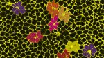
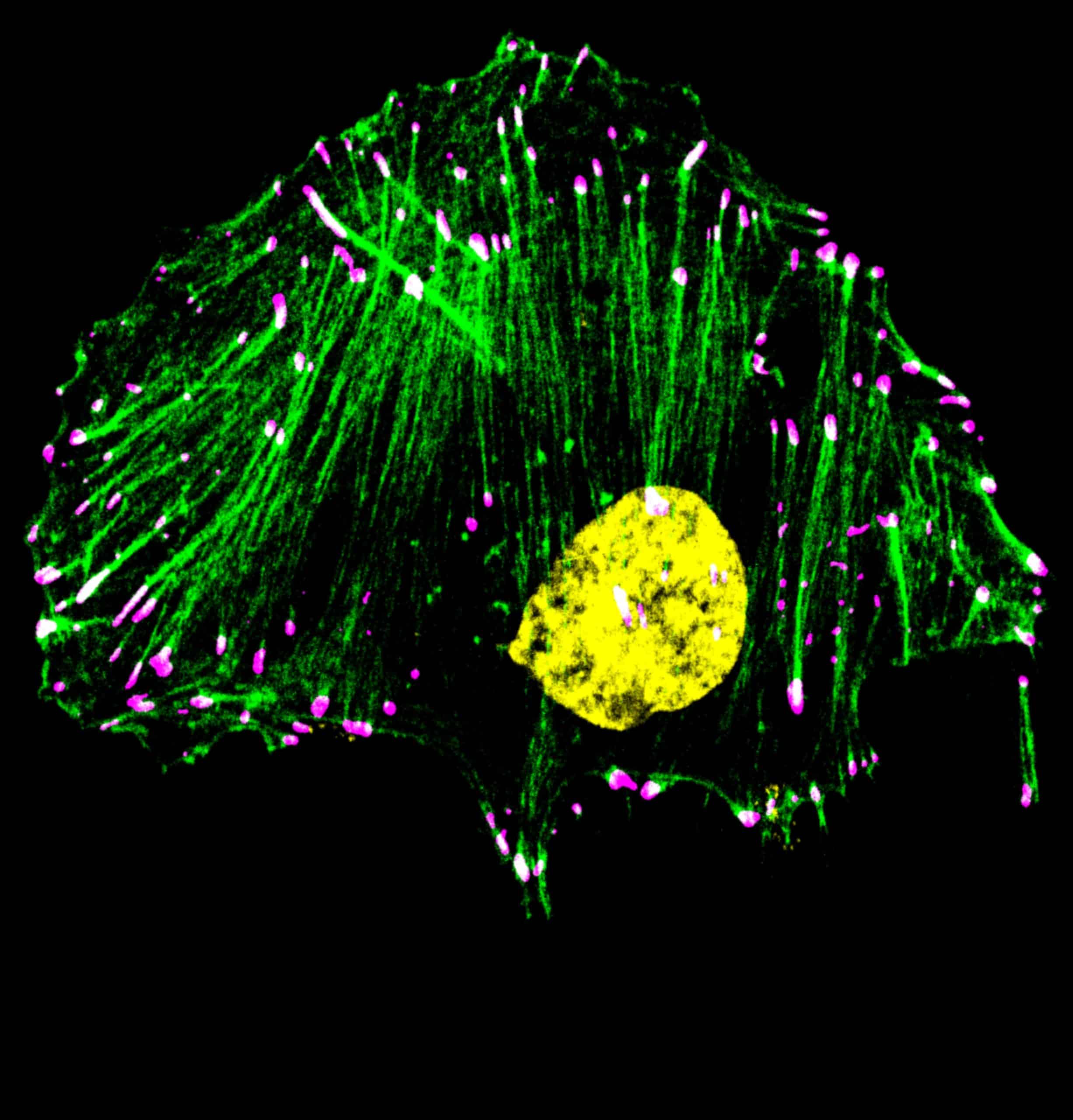
Our featured image, acquired by Manomay Pawar, depicts MDA-MB-231 triple negative breast cancer cells treated with drug Bemcentinib (R428), a small molecule inhibitor of AXL (a receptor tyrosine kinase) and labelled with phalloidin (actin;green), anti-Paxillin antibody to mark focal adhesions (magenta) and DAPI (DNA;yellow). The white coloured puncta represent colocalisation of paxillin with the ends

Microscopy preprints: applications in biology

Posted by , on 15 July 2026

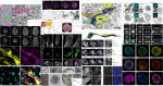
Here is a curated selection of preprints published recently. In this post, we share preprints that use microscopy tools to answer questions in biology.

Compartmentalized Cytoplasmic Tradewinds

Posted by , on 9 July 2026

Cathy Galbraith and Jim Galbraith share their story of answering how does a cell deliver a soluble protein — untethered to any motor, riding on no track — to the right place at the right time? They used six different imaging approaches to discover hidden cytoplasmic compartments and tradewinds that were hiding in plain sight.

Featured image with Joan Roncero Carol

Posted by , on 3 July 2026

Our featured image, acquired by Joan Roncero Carol, shows two mouse preimplantation blastocysts at E4.5 showing immunofluorescence staining against LAMP1 to visualize lysosomal distribution at this developmental stage, together with phalloidin staining to clearly outline cell membranes.

Living Architectures

Posted by , on 30 June 2026

To mark the 40th anniversary of the Musée d’Orsay, the CytoMorpho laboratory was invited to create a performance based on a series of experiments conducted for the occasion. The images produced for this show were real. There were no digital simulations or generative artificial intelligence. They were series of true photographs taken with microscopes.

FocalPlane features... cell migration recordings with Juan Manuel Garcia Arcos, Yohalie Kalukula & Daniel J. Cohen

Posted by , on 18 June 2026

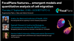
In June, we hosted the first webinar in our new series on emergent models and quantitative analysis of cell migration, hosted by Pablo J. Sáez and Valeria Venturini. We heard talks from Juan Manuel García-Arcos, Yohalie Kalukula and Daniel J. Cohen.

Imaging spotlight: Cryo-ExM reveals the architecture of the human cytotoxic immunological synapse

Posted by , on 15 June 2026

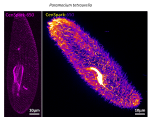
In this behind the paper story, Benita Wolf and colleagues share their research using cryo-expansion microscopy (cryo-ExM) to map the architecture of the human cytotoxic immunological synapse.

Microscopy preprints: applications in biology

Posted by , on 29 May 2026

Here is a curated selection of preprints published recently. In this post, we share preprints that use microscopy tools to answer questions in biology.

New webinar series on cell migration

Posted by , on 29 May 2026

We are excited to announce a new quarterly webinar series on cell migration. In our first webinar, we'll hear talks from Juan Manuel Garcia Arcos, Yohalie Kalukula and Daniel J. Cohen.