

Imaging spotlight: CenSpark
Posted by FocalPlane, on 24 August 2026
In this Imaging spotlight, we hear about CenSpark, a small molecule probe for live imaging of centrioles, from Cédric Pourroy, Georgios Hatzopoulos, Pierre Gönczy and team.
Can you briefly describe the development of CenSpark and what it does?
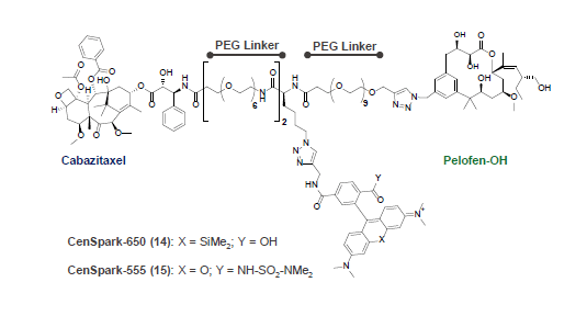
CenSpark was born from a glaring gap: unlike many organelles, centrioles had no dedicated small-molecule probe for selective live visualization. Centrioles have a very distinctive microtubule architecture — nine triplets, each built from one complete A-microtubule and two incomplete B- and C-microtubules. We realized that this triplet arrangement, and the equivalent doublet arrangement in cilia, creates a geometry found nowhere else in the cell: a binding site on the inside of the B-microtubule lumen sits across from a binding site on the outside of the A-microtubule, roughly 44 Å away, close enough to be bridged by a flexible linker.
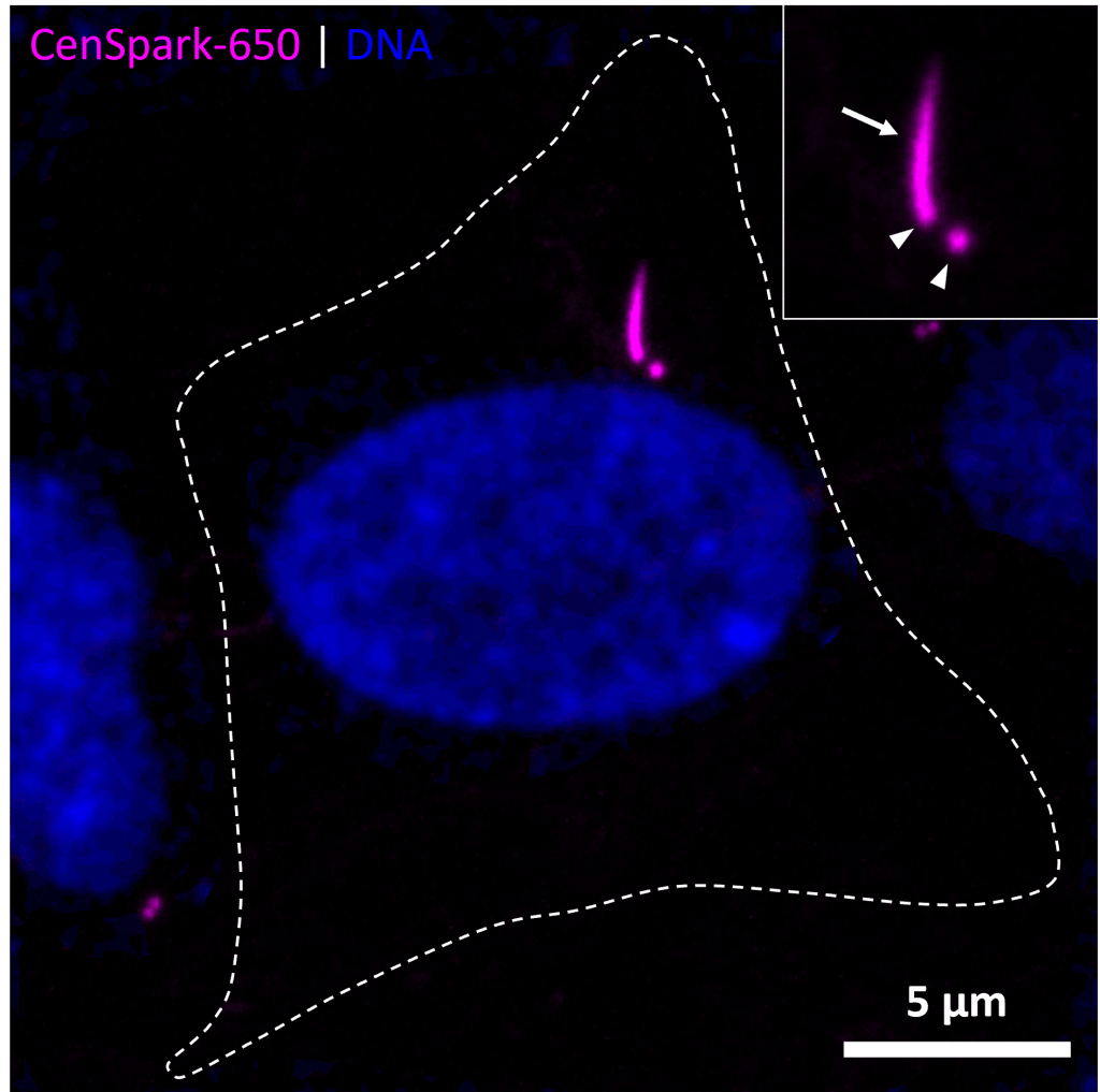
We exploited this by unique geometry by designing a dual-ligand probe that occupies both sites at once: cabazitaxel, which binds in the inside, connected through polyethylene glycol (PEG) linkers to Pelofen, a peloruside A-derived ligand that binds on the outside, with a fluorophore (SiR or MaP555) incorporated into the scaffold. After screening a library of linker lengths and fluorophore positions, we arrived at CenSpark — available as CenSpark-650 (SiR) or CenSpark-555 (MaP555) — the first fluorescent probe that selectively labels centrioles and cilia in living cells, with no genetic manipulation required.
What models has CenSpark been used in so far, and do you anticipate any systems where it won’t work?
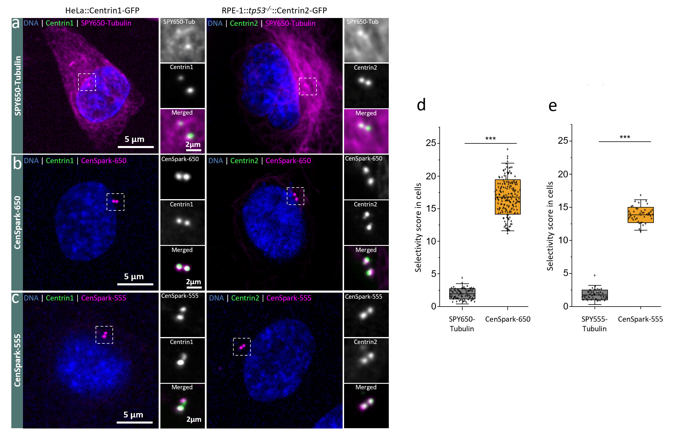
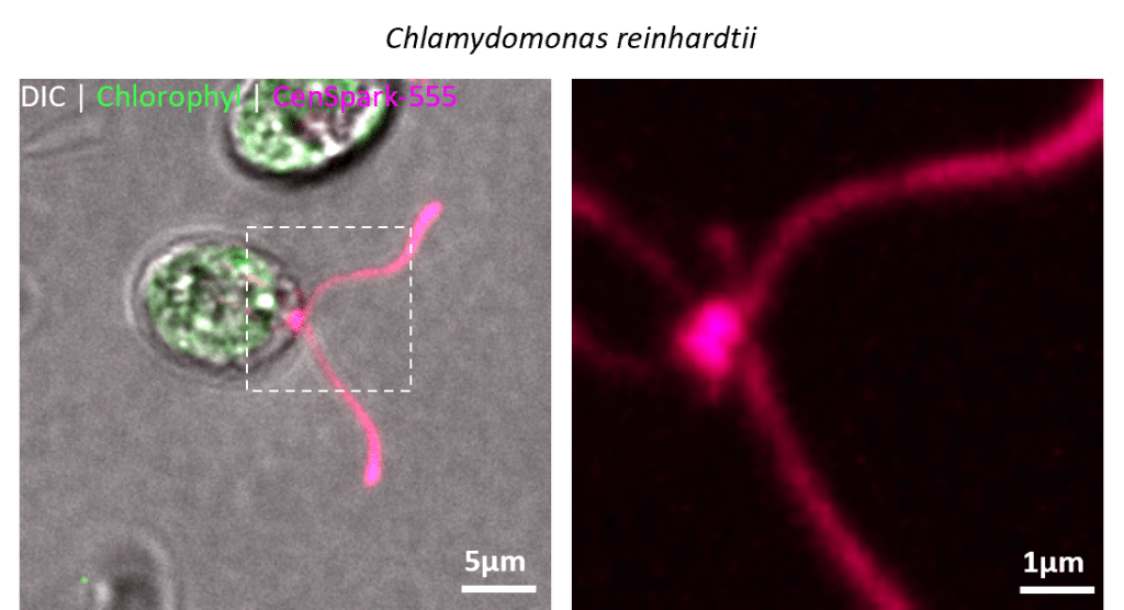
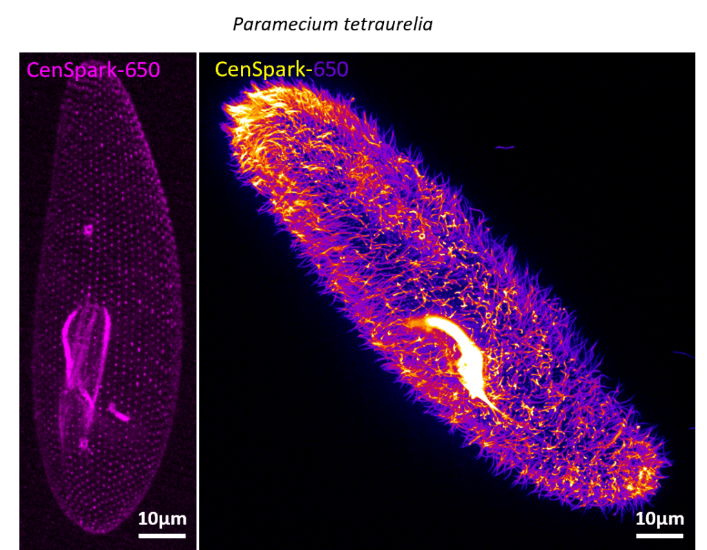
We first validated CenSpark’s selectivity across human cell lines — RPE-1, HeLa and U-2 OS — as well as in primary dermal fibroblasts and chimeric antigen receptor (CAR)-T cells, which are not trivial to engineer genetically. We also tested CenSpark across the range of specimens we work with in the lab: the green alga Chlamydomonas reinhardtii, the ciliate Paramecium multimicronucleatum, Naegleria gruberi, zebrafish (Danio rerio) and Drosophila melanogaster S2 cells. In each case CenSpark labelled centrioles and/or cilia with the expected selectivity.
The one system where CenSpark failed initially was Drosophila spermatocytes imaged ex vivo, where the local membrane composition blocked probe entry. A brief treatment with a mild surfactant was enough to permeabilize the membrane and enable staining. Therefore, our expectation is that CenSpark might not work in systems with unusually impermeable membranes or cell walls, in which cases a short permeabilization step, or else, injection, is likely to solve the problem, as it did for us.
Do you have any tips or tricks for optimising labelling with CenSpark?
CenSpark is simple to use: add it to the medium of your choice at the desired concentration and incubate; no transfection, no cloning, no waiting for expression. In our hands, incubation at 100–500 nM for an hour, followed by a few washes, works well for most human cell lines. As with any live-imaging probe, though, the optimal concentration and incubation time can vary between specimens, so if you’re using CenSpark in a system we haven’t already characterized, we recommend running a pilot titration first.
One trick worth flagging: shorter incubation times and lower probe concentrations actually improve the centriole-to-cytoplasm selectivity, at some cost to overall signal intensity, so there’s a trade-off depending on whether you need maximum contrast or maximum brightness. We also recommend always preparing CenSpark fresh from the DMSO stock into your imaging medium rather than pre-diluting and storing it.
What are the prospects for further development?
The dual-ligand design leaves a lot of room to grow. The fluorophore position in CenSpark can be swapped for a bioorthogonal click handle, which would let researchers attach essentially any warhead of choice after labelling — oligonucleotides for DNA-PAINT super-resolution imaging, for instance. We are also exploring amine-functionalized versions to make CenSpark compatible with expansion microscopy, which typically requires an anchoring handle to covalently link the fluorophore into the swellable gel.
Where can people find more information?
The full study — including all synthesis and validation details — is published open access in Nature Chemical Biology: Pourroy et al., 2026. CenSpark-650 is also available commercially through Spirochrome. We’re always happy to be contacted directly with questions, too (cedric.pourroy@epfl.ch).