Advertisement

Displaying posts with the tag: is_archive

Featured image with Anthony Dornan and Fara van der Schans

Posted by , on 28 February 2025

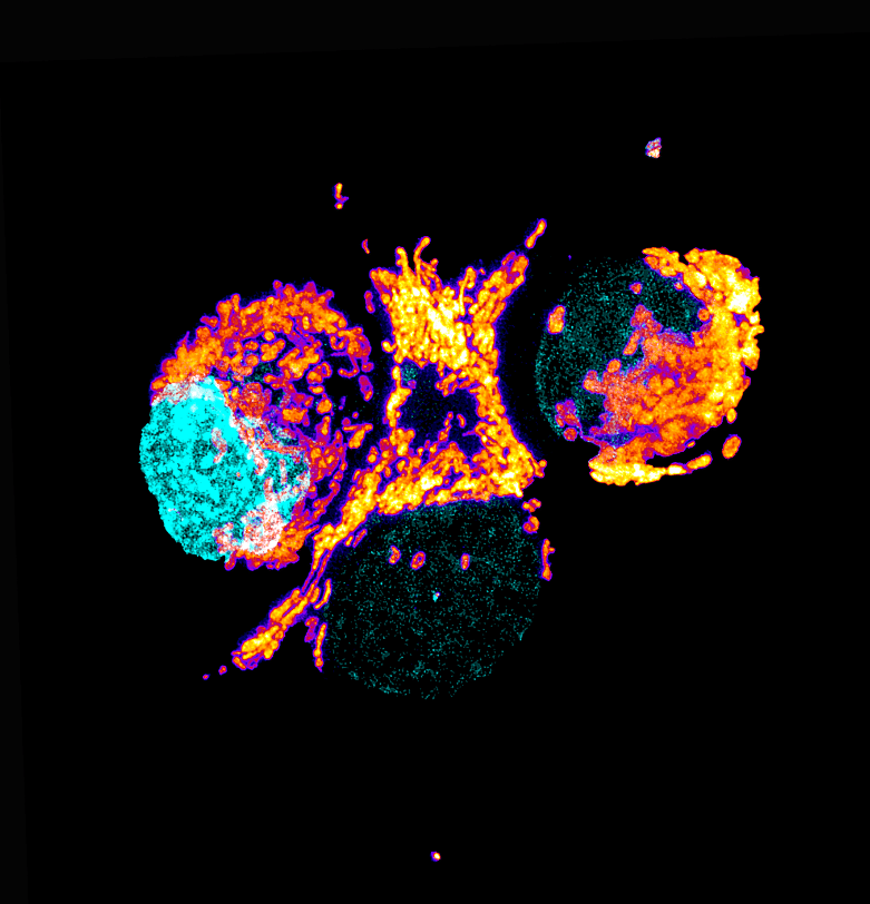
Our featured image, acquired by Anthony Dornan and Fara van der Schans, showcases the perinuclear distribution of mitochondria in the highly respiratory human pancreatic ductal adenocarcinoma (PDAC) tumour cells (Panc-1) stained with the mitochondrial stain TMRE (thermal LUT), the mitochondria are associated with the nuclei, stained with DAPI (cyan). The live cell image is a maximum intensity Z-projection generated

Education Strategies of Imaging Cores

Posted by , on 1 April 2024

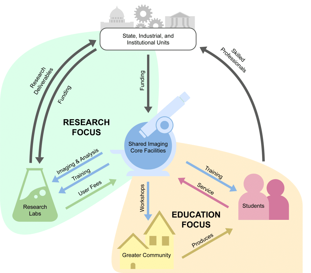
Over the past two decades, high profile research institutions have come to rely on distinct core facilities for much of their data and image analysis. The benefits of the newest technologies are often softened by the costs of purchasing and maintaining such equipment for regular use. For microscopy and imaging, it only made sense that

Imaging with IMAG’IC, the photonic imaging core-facility of Institut Cochin, Paris, France.

Posted by , on 20 February 2024

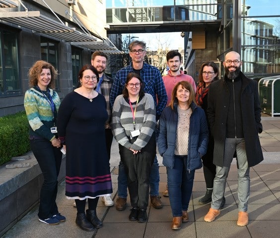
In our ‘Imaging with…’ blog post, we meet the staff of IMAG’IC, the photonic imaging core-facility of Institut Cochin, Paris, France.

Imaging with The University Imaging Centers, a Nikon Center of Excellence, at the University of Minnesota – Twin Cities

Posted by , on 25 January 2024

In our ‘Imaging with…’ blog post, we meet the staff of  The University Imaging Centers, a Nikon Center of Excellence, at the University of Minnesota – Twin Cities.

Enhancing Global Access: interview with CZI grantee Mark Scimone

Posted by , on 24 January 2024

A NEW PROTOTYPE FOR IMAGING CORE FACILITIES As his project for the Cycle 1 imaging scientist call, Mark Scimone proposed a new prototype of what the standard imaging core facility could be. This includes facilitating collaborations between vendors, researchers and cores; generating structured microscopy and optics education courses which also introduce new research technologies; and recruiting new

Enhancing Global Access: interview with CZI grantee Leonel Malacrida

Posted by , on 24 January 2024

DEVELOPING AN ADVANCED BIOIMAGING CORE IN LATIN AMERICA Leonel Malacrida is an Associate Professor at the Pathophysiology Department at Universidad de la Republica in Uruguay. He has recently established the Advanced Bioimaging Unit (UBA for its acronym in Spanish), a joint initiative between Institut Pasteur Montevideo and Universidad de la Republica. The UBA aims to

Enhancing Global Access: interview with CZI grantee Kerry Thompson

Posted by , on 24 January 2024

DEVELOPING A NATIONAL CENTER FOR DOCTORAL TRAINING IN MICROSCOPY Kerry Thompson is an Anatomist and Microscopist in the College of Medicine, Nursing and Health Sciences at the University of Galway in Ireland. Through her project, she aims to establish a National Center for training in microscopy, imaging and related technologies. She is working towards generating

Imaging with the Instituto Gulbenkian de Ciência’s Electron Microscopy Facility

Posted by , on 14 December 2023

In our ‘Imaging with…’ blog post, we meet the staff of the Electron Microscopy (EM) facility at the Instituto Gulbenkian de Ciência, Lisbon, Portugal.

Imaging with the Microscopy and BioImaging Facility at the Leloir Institute Foundation, Buenos Aires, Argentina.

Posted by , on 9 November 2023

In our ‘Imaging with…’ blog post, we meet the staff of the Microscopy and BioImaging Facility at the Leloir Institute Foundation, Buenos Aires.

Light Microscopy Specialist at Virginia Tech

Posted by , on 21 September 2023

The Fralin Life Sciences Institute (FLSI) at Virginia Tech is looking for a highly motivated individual to lead the development of a light microscopy imaging core facility, serve as its manager, and support research groups with microscopy needs on Virginia Tech’s Blacksburg campus. Application review will start on Oct 13th. The Light Microscopy Specialist will