Living Architectures
Posted by FocalPlane, on 30 June 2026
By Léa Blanc, Louise Bonnemay, Simona Buracco, Alice Cantat, Alexandra Colin, Jérémie Gaillard, Christophe Guérin, Laetitia Kurzawa, Nevena Morel, Anne-Betty N’Diaye, Elisa Paulin, Alfredo Sciortino, Flora Silberzan, Bhagyanath Suresh, Clothilde Utzschneider, Benoit Vianay, Laurent Blanchoin & Manuel Théry.
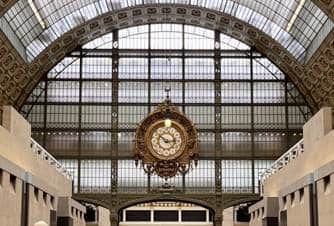
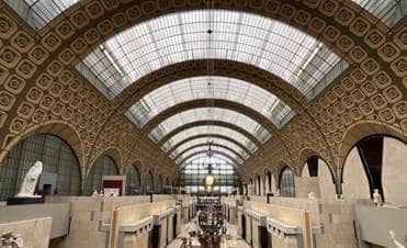
At the musée d’Orsay, January 2026
CytoMorpho Lab, CEA / CNRS, UMR8231, UMR5168, Université Paris Sciences et Lettres, Université Grenoble-Alpes.
To mark the 40th anniversary of the Musée d’Orsay, the CytoMorpho laboratory was invited to create a performance based on a series of experiments conducted for the occasion. The images produced for this show were real. There were no digital simulations or generative artificial intelligence. They were series of true photographs taken with microscopes. The experiments were conducted by members of the CytoMorpho laboratory in Paris and Grenoble. The young students and their supervisors spent several months adapting their everyday experiments to the scenario of the show in order to highlight the questions that drive their research.
The chosen scenario discusses cell morphology and how cells adapt their shape to their environment. In addition to shape, it emphasizes the internal architecture of cells, which also adapts to the environment. It highlights that what is alive is not the cell itself, but the interaction between the cell and its environment. It is this coupling that has allowed life to persist for billions of years.
The scenario emphasizes that living organisms are constantly renewing themselves and that it is thanks to this characteristic that they are able to adapt. Not only do their cells get replaced, but the internal components of the cells are also continuously renewed. The internal architecture of cells consists of small filaments. These filaments are interconnected and form large networks. Some filaments assemble, others disassemble. Some replace others, and gradually the entire network renews itself while maintaining its structure. Thanks to this dynamic, the network can grow on one side or contract on another, thereby changing the shape of the cell. Images from the experiments suggest that for a cell, the time that matters is not the second or the minute, but the time it takes to renew itself. It defines the intrinsic timescale at which the cell can sense and adapt to its environment.
Finally, the scenario explores the limits of the plasticity of living organisms. The performance first illustrates the process by which cells change shape and reorganize their filament networks, revealing a remarkable coherence between the interior and exterior of the cell, and between the structure of its filament network and the architecture of the museum. The filaments change orientation depending on the arrangement of the beams or the spacing of the museum’s stones. This impressive plasticity raises the question of the limits of this process. The latest experiments push the cells to their limits. To what extent can the cell construct itself in the image of the museum? Are certain shapes impossible? Are certain sizes beyond reach? Can the cell cover the entire museum?
Methodology
For the past twenty years, the CytoMorpho laboratory has been developing and using lithography techniques derived from microelectronics to print micrometer-scale patterns and control cell shape. The lab also uses the same methods to study the growth and organization of the filaments that make up the cell skeleton.
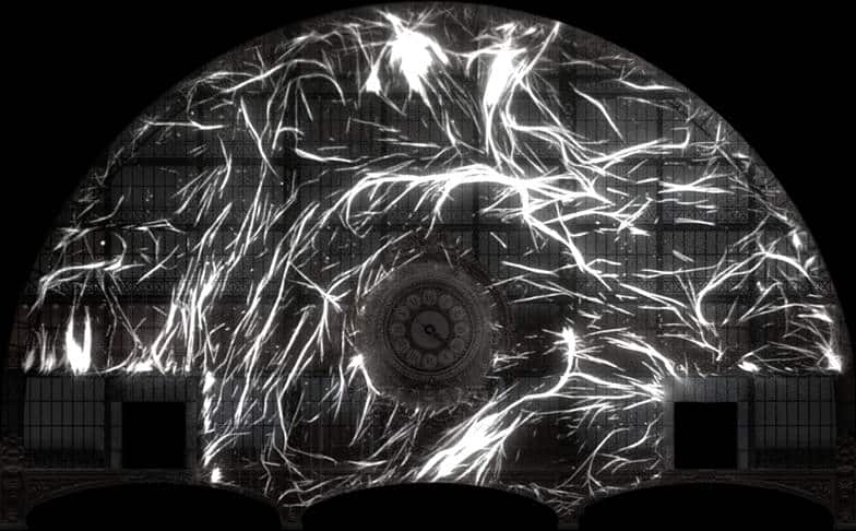
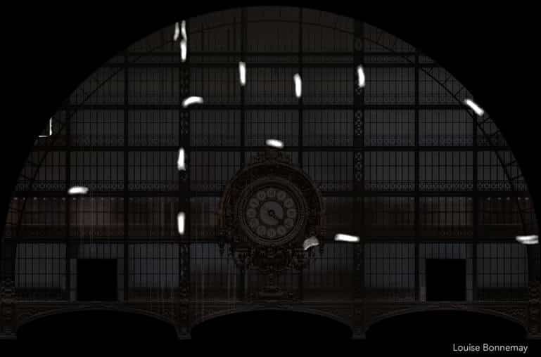
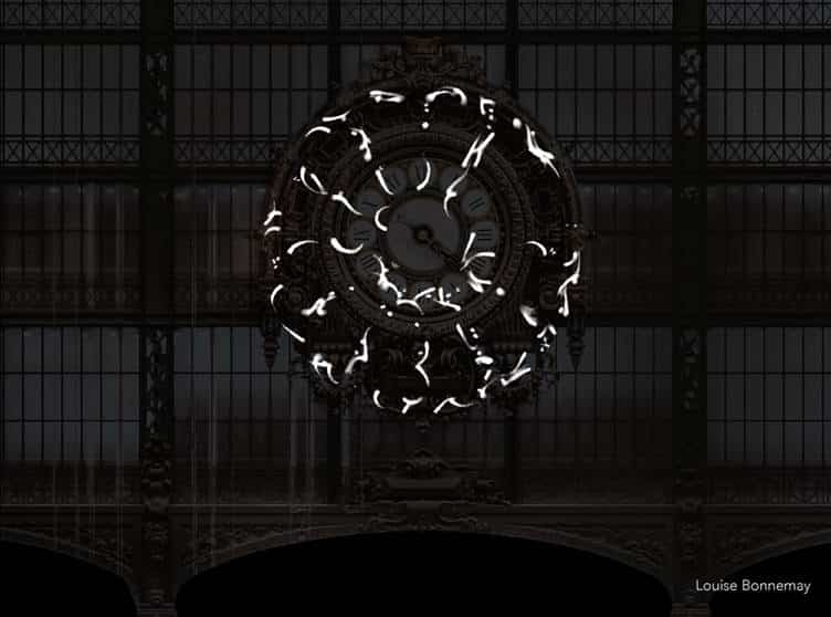
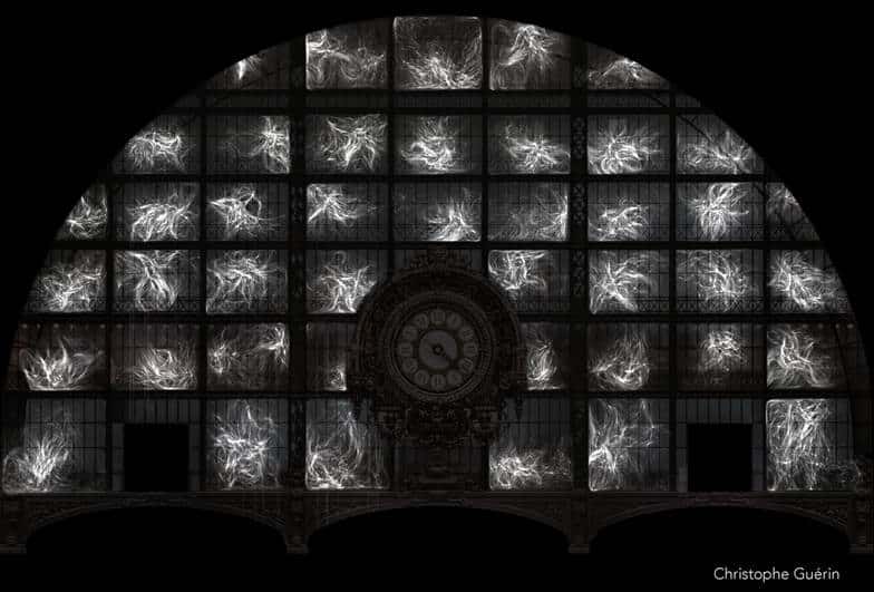
In this project at the Musée d’Orsay, the architectural elements of the façade, where the clock is located, and of the nave that crowns the museum have been miniaturized to the cellular scale using UV light.

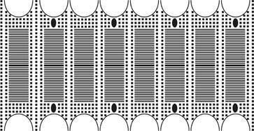
The light was structured using optical masks or arrays of micromirrors to project the desired patterns onto glass slides. These patterns were then used to attach various proteins capable of promoting cell adhesion or filament growth. The cells thus attached themselves to patterns that represented the museum’s architecture. The filaments grew from the same type of patterns. The behavior of the cells and filaments was then filmed for hours, even days, using various microscopes equipped with highly sensitive digital cameras.
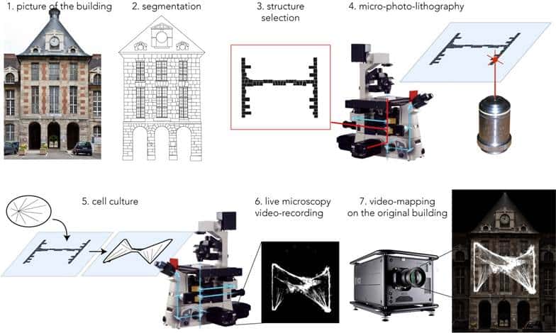
During the performance, the resulting films were projected using powerful video projectors onto the original structures that had been printed on the glass slides. The audience was thus able to see the cells and filaments conforming to the building’s structures. These unprecedented images led them to wonder about the mechanisms that give living architecture this plasticity.
An orchestra provided musical accompaniment for the film screenings, and an actress read a poetic text that guided the audience in their reflections and daydreams.
Introduction, the filaments of cell skeleton
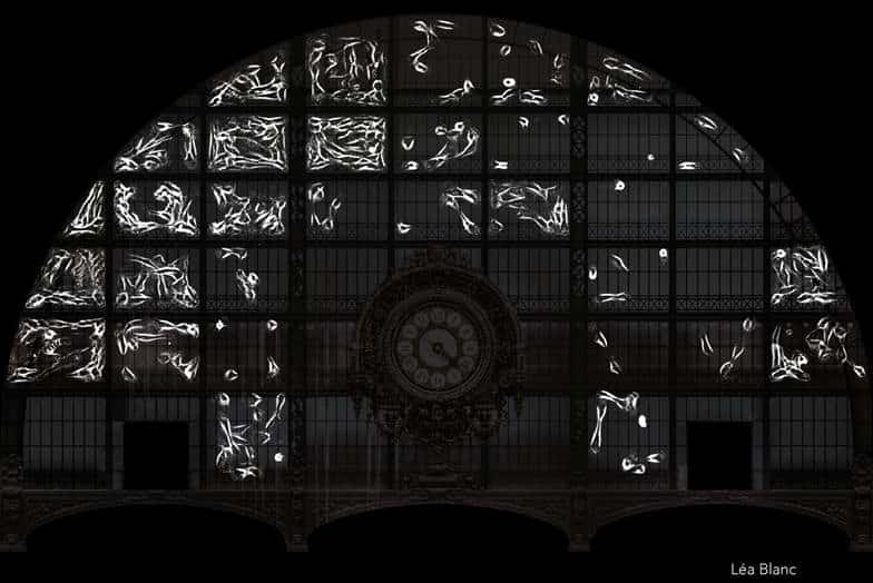
The opening images of the show illustrate the emergence of basic structures by depicting the formation and growth of filaments. As they appear on the museum ceiling, we see that these filaments are dynamic, alternating lengthening and shortening. This dynamic illustrates, in a simple way, that life destroys as much as it builds.
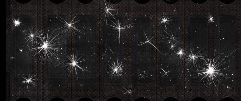
In these experiments, the filaments shown are not found in living cells. They are assembled from purified proteins. Indeed, the researchers of the team study not only the cells but also the intrinsic properties of the filaments that make up their cytoskeleton. To do this, they break down the cells and isolate only the proteins involved in filament assembly. These proteins are then recombined in precise proportions to study how these filaments assemble and form networks. The images thus show biological filaments, isolated from cells, revealing their most fundamental properties.
Part One: The Emergence of Order and the Genesis of Forms
This series of experiments illustrates how living organisms create order by forming patterns. When added to the mixture, certain proteins, the so-called molecular motors, exert forces on the filaments and cause them to move. When there are few filaments, their trajectories appear random, but as their number increases, they align themselves until they move in a completely coordinated manner. Axes of symmetry emerge in their movements, and the erratic paths become oriented in space.
If we then add other molecular motors moving in the opposite direction, the opposing forces balance each other out and the filaments come to a standstill. As they align, the filaments trace boundaries that define emerging shapes. These shapes are not static; they are “active”: they consume energy to maintain themselves. If the balance of forces is altered, they can deform. If it is broken, they disappear and disorder takes over again.
Filaments can also generate forces by lengthening. This allows them to push the object they are growing against. This is the mechanism that causes cell membranes to deform. In these experiments, the filaments push beads, which can then explore the space.
At high densities, another phenomenon that generates shape and order emerges: the beads arrange themselves into a regular pattern. Spontaneously, they align and position themselves at equal distances from one another, forming a sort of crystal. This surprising phenomenon has never been described before and was discovered during experiments conducted for the event.
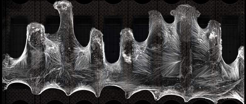
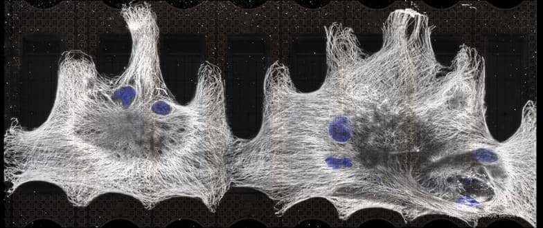
Part two: Integration of the Museum’s Architecture
The cells attach onto the museum’s beams and windows. They explore the entire surface and attempt to cover it. Indeed, the cells do not have a predetermined shape. They change shape depending on the structures they encounter. In doing so, they conform to the museum’s architecture.
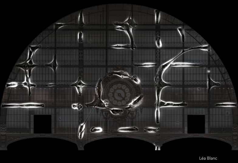
When images are captured at faster time scales, it becomes apparent that every movement is accompanied by a complete reorganization of the filaments within the cell. The internal network is constantly being completely broken down and rebuilt. If the cell membrane comes into contact with vertical beams, the filaments align with them. If, as it moves, the cell encounters other perpendicular beams, the filaments change orientation and guide the cell in this new direction. Everywhere, filaments are broken down so that new ones can form on the structures the cells encounter. This mechanism of constant reconstruction allows the filament network to replicate the architecture encountered outside the cells within the cells themselves. The cells internalize the image of their environment. Thus, the cells can sense and continuously adapt to changes in their environment.
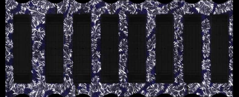
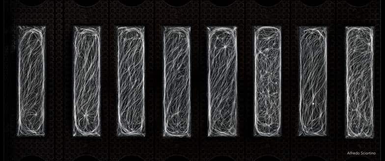
1. actin filaments in human cells migrating on adhesive micropatterns with the shape of the metal beams of the clock facade (Elena Rossetti, Laetitia Kurzawa)
2.actin filaments in a single human cell covering an adhesive micropatterns with the shape of the clock facade (Laetitia Kurzawa)
3. actin filaments in a single human cell covering an adhesive micropatterns with the shape of the horizontal metal beams the clock facade (Laetitia Kurzawa)
4. actin filaments in a single human cell covering an adhesive micropatterns with the shape of the clock facade (Laetitia Kurzawa).

The films reveal that within the cell, everything that has been built must be completely broken down. The breakdown must be total so that all components can be reused. This complete recycling and constant renewal are hallmarks of life.
This impressive ballet inside each cell requires perfect coordination. If the filaments grow slightly faster than they are broken down, the cell will expand and risk tearing its membrane. Conversely, if the filaments are broken down a little too quickly, the network will gradually shrink and the cell risks collapsing in on itself. How is this balance achieved? The survival of all living systems relies on it. This question is as simple as it is fundamental.
Part Three: Building a Living Structure
As important as they may be, some questions are too difficult to solve in contexts as complex as the interior of a cell. Hundreds of thousands of different proteins are involved in cross-reactions. It’s unsolvable.
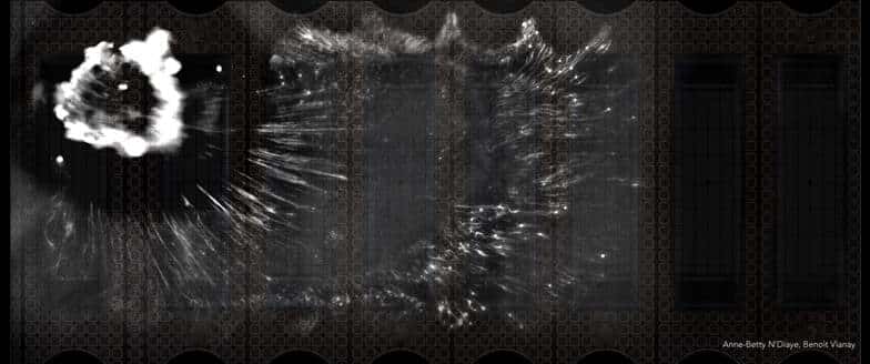
The team therefore attempted to construct a living architecture using a minimal number of components. Microstructured scaffolds were used to guide the assembly of filament networks that were forced to grow from the museum’s architectural elements.
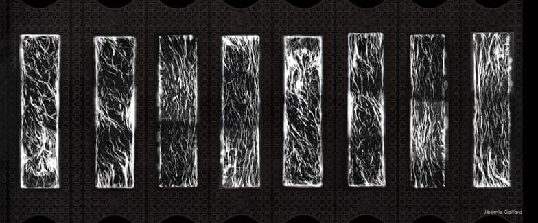
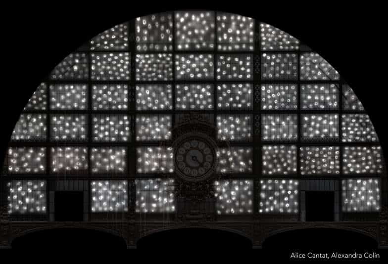
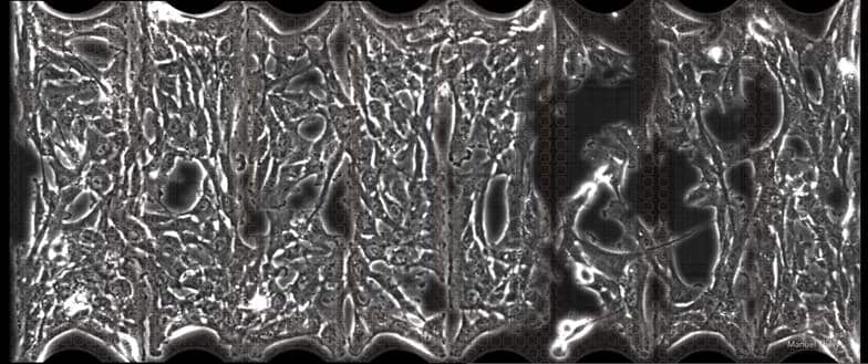
On the museum’s windows, the first filaments appear. Each filament allows for the assembly of another filament, so their number grows exponentially. Soon the windows are covered with filaments, just as is the case along cell membranes. The filaments align alongside one another and form arabesques with regular curves.
By modifying the microstructure in contact with the purified filaments, it is possible to control the starting point of their growth. When growing from the stones, the filaments align themselves and form a regular mesh that connects neighboring stones. When growing from the beams, the filaments form perpendicular cables.
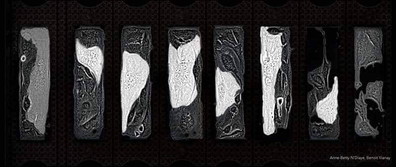
1. growth of actin filaments starting from micropatterns with the shape of the horizontal metal beams of the clock facade (Christophe Guérin)
2. growth of actin filaments starting from micropatterns with the shape of the vertical metal beams of the clock facade (Nevena Morel)
3. growth of actin filaments on micropatterns with the shape of the arches of the nave ceiling (Christophe Guérin)
4. growth of actin filaments on micropatterns with the shape of windows of the nave ceiling (Nevena Morel).
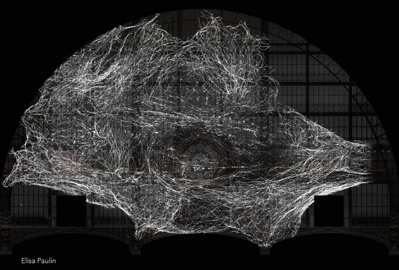
But these beautiful networks are static and bear little resemblance to the dynamic networks observed in cells.
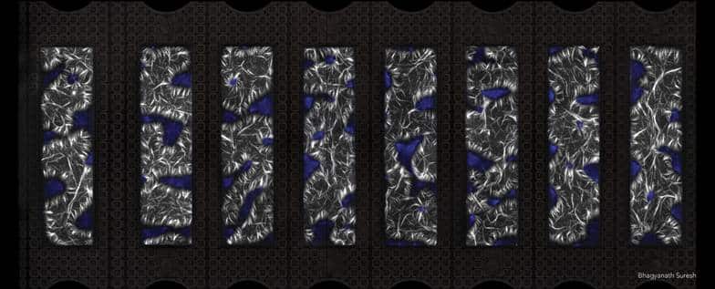
When additional proteins—molecular motors—are added to the mixture, the filaments align to form cables that contract. The entire network then comes to life with movements resembling those observed in cells. But at the end of the contraction, the filaments are destroyed. The desired balance between construction and destruction is not achieved, and the entire structure that had been built collapses abruptly.
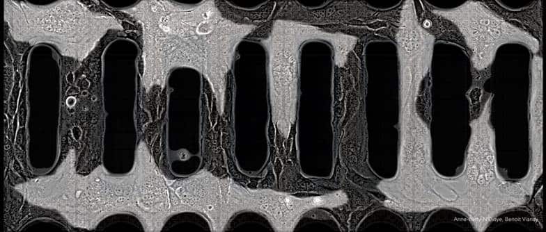
1. contraction by myosins of a network of actin filaments assembled from micropatterns with the shape of the horizontal metal beams of the clock facade
2. contraction by myosins of a network of actin filaments assembled from micropatterns with the shape of the vertical metal beams of the clock facade
3. contraction by myosins of a network of actin filaments assembled from micropatterns with the shape of the vertical metal beams of the clock facade
4. contraction by myosins of a network of actin filaments assembled from micropatterns with the shape of the arches of the nave ceiling
5. contraction by myosins of a network of actin filaments assembled from micropatterns with the shape of the arches of the nave ceiling
6. contraction and disassembly by myosins of a network of actin filaments assembled from micropatterns with the shape of the arches of the nave ceiling.
Under certain conditions, the structure’s dynamics manage to strike a balance between contraction, destruction, and reassembly. But these equilibria are precarious, and the logic behind their robustness within cells eludes us.
As surprising as it may seem, all these impressive structures, which are the result of billions of years of evolution, remain extremely fragile. Perhaps that is, in fact, the core of the mechanism: being fragile, on the verge of breaking, in order to be able to erase everything, rebuild everything, and thus constantly explore.
Part Four: Exploring the Limits
We have constructed filament networks ten to a hundred times larger than the normal size of cells. Perhaps no equilibrium is possible at these scales. Perhaps we are approaching the stability limit of living architectures. We do not even know if such an equilibrium would be possible in cells of such a size.
The animal cells we used for this project measure nearly thirty micrometers. Since the micro-museum is a thousand micrometers long, the cells cover only a small portion of it. Without even raising the fundamental question of the intrinsic limits of the size of living units, we might wonder what would happen to a cell if we forced it to be a hundred times larger than its normal size. Such a cell could cover the entire museum, but wouldn’t it collapse as well?
We therefore established a new protocol to create giant cells and observe the dynamics and equilibrium of their filament network. We forced neighboring cells to fuse together using viral proteins that weaken membrane permeability. Very large cells, resulting from the fusion of several cells, manage to cover a large portion of the museum but not its entirety.
By forcing a few very large cells to fuse again together, we end up with giant cells containing dozens of nuclei. These monsters nearly cover the entire museum, and the structure of their filament network bears no resemblance to what we’re used to seeing. The entire network seems to have lost its coherence.
But these monsters are not viable. A few hours after they form, they collapse and die. Their existence was therefore limited to this unique experiment.
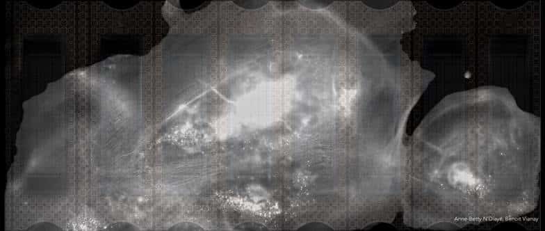
We were therefore unable to cover the entire one-millimeter-long micro-museum with a living system. Neither the structures reconstructed ex vivo nor the giant cells were able to maintain themselves at such a small scale. After these few experiments, we cannot draw any conclusions about the generality of this phenomenon, but it is possible that we have opened a window onto the stability limit of the dynamic filament structures that underpin cell shape.
Conclusion
All of these experiments illustrate that it is by constantly breaking down that living systems are able to rebuild themselves by adapting to changes in their surroundings. The bonds between biological components are so fragile that they can be easily broken and rebuilt, which gives living structures great plasticity. This plasticity allows cells to constantly adapt to their environment. Unlike inert materials, in which the strength of the bonds allows them to resist and endure, it is the fragility of these bonds that allows living systems to constantly reinvent themselves. But their plasticity is also their weakness. If the balance between destruction and construction is not perfect, the system cannot hold together and dies quickly.
Cells thus use their internal dynamics to explore every possibility, without goal or intention, simply because it is possible. Anything too unstable will collapse and make way for other attempts. Cells therefore spend their time playing with the limits of their stability. Most of them will die as a result. We are today the product of an evolution paved by the death of trillions upon trillions of cells. These adventurers did not attempt to achieve a specific goal that would have made them more robust or more efficient. They tried everything made possible by their internal dynamics.
Acknowledgements
The experiments were conducted at Institut de Recherche Interdisciplinaire de Grenoble (IRIG) and the Ecole Supérieure de Physique et Chimie Industrielle de la Ville de Paris (ESPCI ParisTech) with support of the Chair of Excellence in Biology and Health (ANR-23-CHBS-0013), as part of the France 2030 program, and with the support of the European Union’s Horizon 2020 research and innovation programme under the Marie Skłodowska-Curie grant agreement No 101108326.
The two-night show at the Musée d’Orsay received support from the metropole du Grand Paris.
 (No Ratings Yet)
Get involved
Create an account or log in to post your story on FocalPlane.
More posts like this


Filter by
- NewsApply
- DiscussionsApply
- How toApply
- ToolsApply
- Case studiesApply
- InterviewsApply
- JobsApply
- EducationApply
- Blog seriesApply
- Volume EMApply
- Latin American Micro..scopistsApply
- Bio-image Analysis w..ith NapariApply
- Imaging with…Apply
- Towards Global Acces..sApply
- Latin America Bioima..gingApply
- From Zero to Qupath ..HeroApply
- Asian Microscopists ..and Cell BiologistsApply
- AIC at HHMI JaneliaApply
- Deep Learning for Bi..o-image analysisApply
- GloBIAS – updates fr..om the communityApply
- WAMBIAN: West Africa.. in FocusApply
- Highlights from Euro..-BioImagingApply
- LSFM seriesApply
- DIY MicroscopyApply
- View all