Advertisement

Displaying posts with the tag: is_archive

Featured image with Md Hashim Reza

Posted by , on 22 November 2024

Our featured image, prepared by Md Hashim Reza, is an Andy Warhol-inspired illustration of a Candida albicans cell, post-expansion and labelled with BodipyTR, highlighting features of internal membrane organisation.

FocalPlane features... Imaging Cell Architecture and Dynamics with Journal of Cell Science

Posted by , on 15 November 2024

Our latest FocalPlane features… webinar was a celebration of the research in Journal of Cell Science’s Special Issue: Imaging Cell Architecture and Dynamics. Our Guest Editors Lucy Collinson and Guillaume Jacquemet hosted the webinar with talks from Md Hashim Reza, Melanie Rug and Vikas Tillu.

Journal of Cell Science Special Issue: Imaging Cell Architecture and Dynamics

Posted by , on 5 November 2024

Journal of Cell Science’s Special Issue on Imaging Cell Architecture and Dynamics is now complete. The original research in this Issue, guest edited by Lucy Collinson and Guillaume Jacquemet, covers a diverse range of imaging methodologies including CLEM, super-resolution imaging, volume EM, high-throughput imaging, FRET, expansion microscopy, mesoscopy and immuno-SEM, as well as showcasing new

FocalPlane features... Imaging Cell Architecture and Dynamics with Journal of Cell Science

Posted by , on 17 October 2024

Our November webinar is a celebration of Journal of Cell Science’s Special Issue: Imaging Cell Architecture and Dynamics. Our Guest Editors, Lucy Collinson and Guillaume Jacquemet, have invited the authors of three of the papers published in this issue to present their research. View the ‘Special Issue: Imaging Cell Architecture and Dynamics’ here. Thursday 14

Featured image with Ciarán Butler Hallissey

Posted by , on 13 September 2024

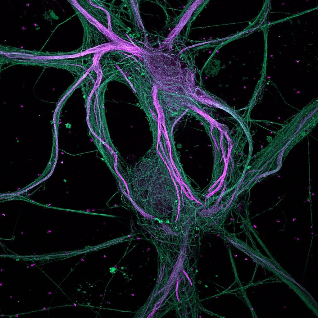
Our featured image, acquired by Ciarán Butler Hallissey, is a maximum-intensity projection of rat hippocampal neurons processed with ultrastructure expansion microscopy (U-ExM) and captured with a spinning disk confocal microscope. Find out more about the image and Ciarán’s research below. More about the image: Expansion microscopy physically increases the size of your sample to improve

Technology Highlights - Expansion Microscopy

Posted by , on 29 August 2023

Interview with Ana Agostinho and Steven Edwards, at the Advanced Light Microscopy facility at SciLifeLab in Sweden. We are today talking about Expansion Microscopy imaging. Please provide a short summary of this type of imaging and list some applications: Ana: My name is Ana Agostinho and I am a staff scientist at the Advanced Light

Advances in Expansion Microscopy

Posted by , on 4 August 2023

Expansion microscopy was introduced in 2015 by Boyden and his team, revolutionizing the way we see biological samples under a microscope1. Taking advantage of the power of physical enlargement, ExM pushes spatial resolution beyond the diffraction limit, allowing us to observe intricate details at previously unimaginable levels. The underlying principle of ExM lies in a