

Advertisement

Displaying posts with the tag: is_archive

vEM reveals the cellular composition of the Blood Nerve Barrier

Posted by , on 11 October 2024

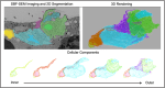
Using Serial Block-Face-SEM6 and AT-SEM, we characterized the cellular composition and interactions along EndoBVs7. Unlike the BBB, we observed that the vasculature within the endoneurium is not completely covered. Upon closer examination of the cells in contact with EndoBVs, we identified three distinct cell types frequently interacting with the vasculature.

Kidney elastic fiber network changes in Marfan's Syndrome

Posted by , on 19 July 2024

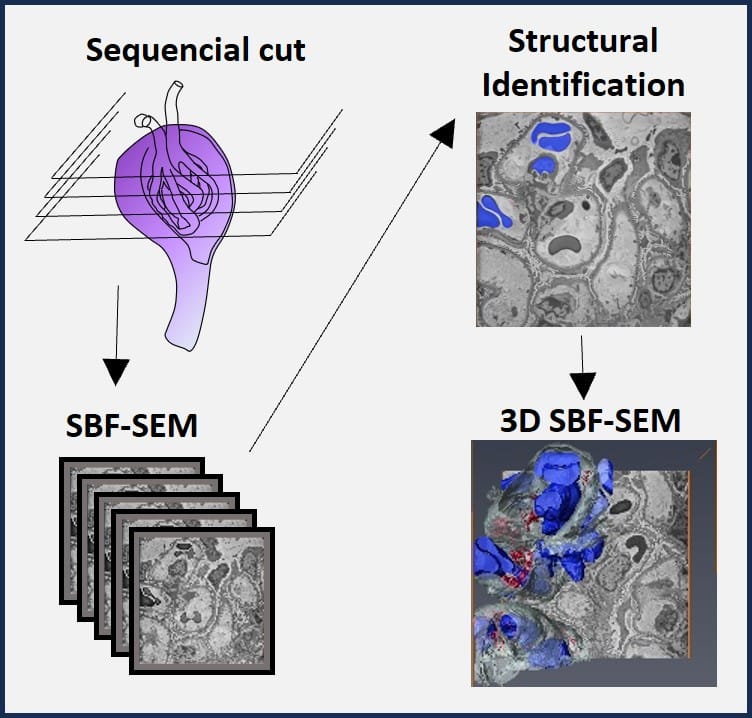
by de Souza RB*, Meek KM#, Lewis PN#, Pereira LV*. *Department of Genetics and Evolutionary Biology, University of São Paulo, Brazil #StructuralbBiophysics Research Group, School of Optometry and Vision Sciences of the United Kingdom, Cardiff University, UK Challenge Marfan’s syndrome (MFS) is an autosomal dominant disorder that affects the connective tissue, resulting from a mutation

Targeted vCLEM of heterogeneous samples

Posted by , on 15 January 2024

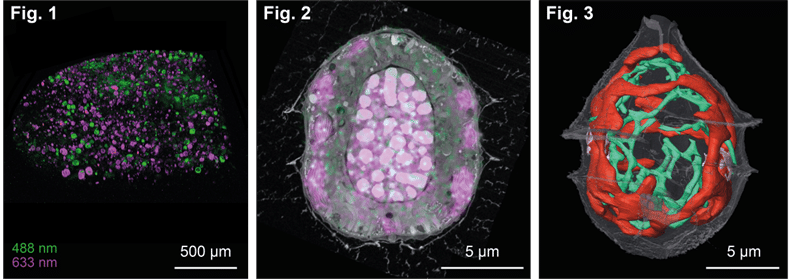
by Karel Mocaer* and Paolo Ronchi# *Schwab Team and #EMCF, EMBL Heidelberg, Germany Challenge Studying cells or events within their biological context (i.e. in tissues, ecosystem etc…) is highly relevant. However, volume EM techniques are limited in the volume that can be acquired. Therefore, targeting approaches are needed to characterize specific regions of interest. This

Deciphering cardiac multiscale organization

Posted by , on 11 December 2023

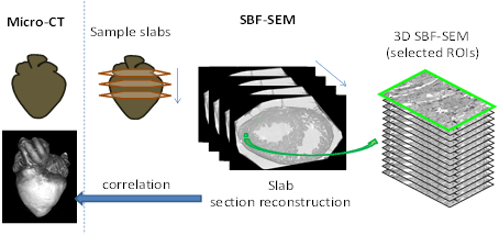
by Sandra Rugonyi and Claudia López Oregon Health & Science University, Portland Oregon, USA Challenge The morphology, microstructure, and ultrastructure of heart tissues is exquisitely organized to optimize cardiac efficiency. In cases of cardiac malformations, known as congenital heart disease (CHD), spatially heterogeneous and abnormal ultrastructural patterns emerge as early as fetal stages that worsen

Unveiling the Marvels of the First-Ever Gordon Research Conference on Volume Electron Microscopy - Capturing Subcellular Architecture from Organelles to Organisms -By Ana Correia and James Thompson

Posted by , on 24 August 2023

We delve into our captivating journey at the first Gordon Research Conference (GRC) on Volume Electron Microscopy (vEM) as early career scientists. Set against the backdrop of sunny Ventura, California, this conference brought together experts in the vEM field from all corners of the globe, creating an intellectual haven for learning, networking, and forging collaborations.

Insights into the leaky Blood Brain Barrier by vEM

Posted by , on 4 August 2023

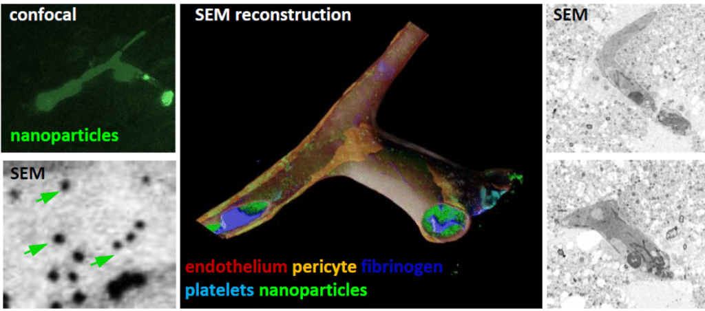
by Martina Schifferer, German Center for Neurodegenerative Diseases, Munich Cluster for Systems Neurology SyNergy, Germany DOI: 10.1242/focalplane.15936 Challenge The Blood Brain Barrier (BBB) controls the exchange between blood and the brain parenchyme. Its structural determinants comprise tight junctions and endothelial vesicles which require resolution at the nanometer scale. In disease like stroke or traumatic brain

Inputs and Outputs of vEM in a Sensory System

Posted by , on 19 May 2023

by Federica Mangione, The Francis Crick Institute, UK DOI: 10.1242/focalplane.14809 Challenge The sense of touch enables organisms to interact with their environment by perceiving physical forces and guiding complex behaviors. Touch sensing is mediated by sensory neurons that innervate the body surface of animals and, together with surrounding cells, form specialized structures known as tactile

Electron microscopy: from the dark ages to a bright future

Posted by , on 21 July 2020

Good sample preparation is, as every microscopist knows, the key to delivering sound results from an imaging experiment. In the digital age, and with the advent of big data, image analysis is also critical to extract meaningful quantitative results from image data. Indeed nowadays, the microscope itself is often the most well-developed and user-friendly part