Microscopy preprints – applications in cell and developmental biology
Posted by FocalPlane, on 30 June 2023
Here is a curated selection of preprints published recently. In this post, we share preprints that use microscopy tools in cell biology and developmental biology.
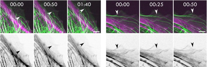
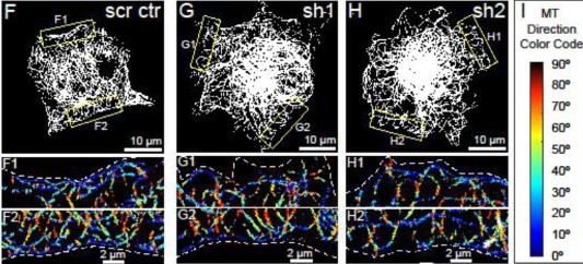
Microtubules under mechanical pressure can breach dense actin networks
Matthieu Gélin, Alexandre Schaeffer, Jérémie Gaillard, Christophe Guérin, Benoit Vianay, Magali Orhant-Prioux, Marcus Braun, Christophe Leterrier, Laurent Blanchoin, Manuel Théry
Multiscale imaging of corneal endothelium damage and effects of Rho Kinase inhibitor application in mouse models of acute ocular hypertension
Zhen Cai, Yang Zhang, Raymond S. Fang, Benjamin Brenner, Junghun Kweon, Cheng Sun, Jeffery Goldberg, Hao F. Zhang
GTPase activating protein DLC1 spatio-temporally regulates Rho signaling
Max Heydasch, Lucien Hinderling, Jakobus van Unen, Maciej Dobrzynski, Olivier Pertz
Two subtypes of GTPase-activating proteins coordinate tip growth and cell size regulation in Physcomitrium patens
Jingtong Ruan, Linyu Lai, Hongxin Ou, Peishan Yi
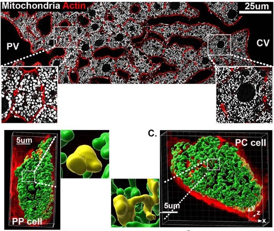
A spatial map of hepatic mitochondria uncovers functional heterogeneity shaped by nutrient-sensing signaling
Sun Woo Sophie Kang, Rory P. Cunningham, Colin B. Miller, Constance M. Cultraro, Jonathan Hernandez, Lisa M. Jenkins, Alexei Lobanov, Maggie Cam, Natalie Porat-Shliom
Self-extinguishing relay waves enable homeostatic control of human neutrophil swarming
Jack Strickland, Deng Pan, Christian Godfrey, Julia S. Kim, Alex Hopke, Maureen Degrange, Bryant Villavicencio, Michael K. Mansour, Christa S. Zerbe, Daniel Irimia, Ariel Amir, Orion D. Weiner
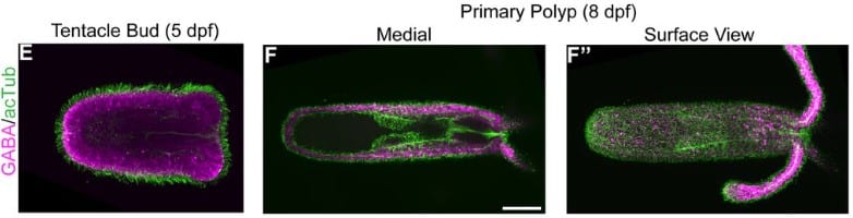
Graded FGF activity patterns distinct cell types within the apical sensory organ of the sea anemone Nematostella vectensis
Keith Z. Sabin, Shiyuan Chen, Eric Hill, Kyle J. Weaver, Jacob Yonker, MaryEllen Kirkman, Bret Redwine, Anna M. L. Klompen, Xia Zhao, Fengli Guo, Cathy McKinney, Jessica L. Dewey, Matthew C. Gibson
The Tubulin Nano-Code: a protofilament-specific pattern of tubulin post-translational modifications regulates ciliary beating mechanics
Gonzalo Alvarez Viar, Nikolai Klena, Fabrizio Martino, Adrian Pascal Nievergelt, Gaia Pigino
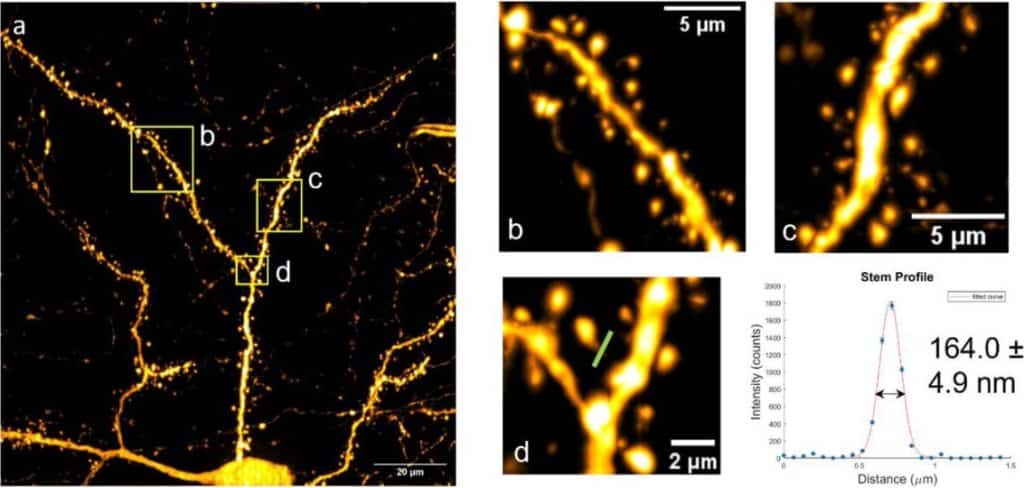
Super-resolution imaging of neuronal structure with structured illumination microscopy
Tristan C. Paul, Karl A. Johnson, Guy M. Hagen
But, what are the cells doing? Image Analysis pipeline to follow single cells in the zebrafish embryo
Arianne Bercowsky-Rama, Olivier F. Venzin, Laurel A. Rohde, Nicolas Chiaruttini, Andrew C. Oates
Content-enriched fluorescence lifetime fluctuation spectroscopy to study bio-molecular condensate formation
Eleonora Perego, Sabrina Zappone, Francesco Castagnetti, Davide Mariani, Erika Vitiello, Jakob Rupert, Elsa Zacco, Gian Gaetano Tartaglia, Irene Bozzoni, Eli Slenders, Giuseppe Vicidomini
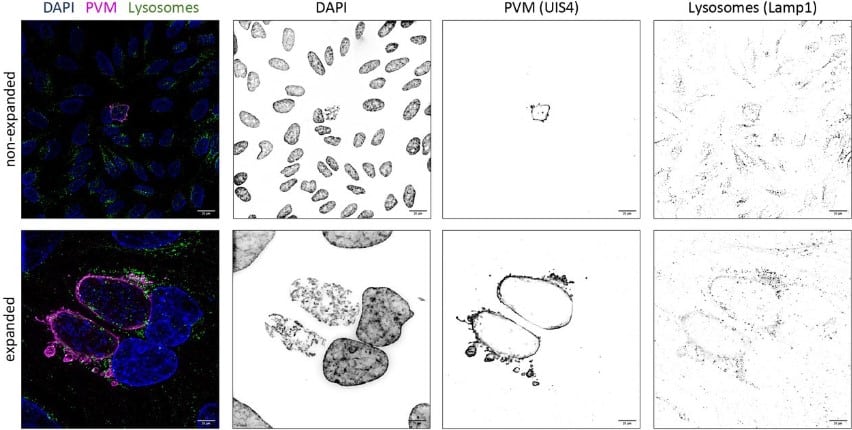
Pre-gelation staining expansion microscopy (PS-ExM) for visualization of the Plasmodium liver stage
Kodzo Atchou, Bianca Manuela Berger, Volker Heussler, Torsten Ochsenreiter
Peroxisomal tail-anchored proteins do not reach peroxisomes via ER, instead mitochondria can be involved
Tamara Somborac, Güleycan Lutfullahoglu Bal, Kaneez Fatima, Helena Vihinen, Anja Paatero, Eija Jokitalo, Ville O Paavilainen, Svetlana Konovalova
Rim aperture of autophagic membranes balances cargo inclusion with vesicle maturation
Oren Shatz, Milana Fraiberg, Alexandra Polyansky, Eyal Shimoni, Tali Dadosh, Sharon Wolf, Zvulun Elazar
Analysis of cortical cell polarity by imaging flow cytometry
Jesper Huitfeld Jespersen, Andras Harazin, Anja Bille Bohn, Anni Christensen, Esben Lorentzen, Anna Lorentzen
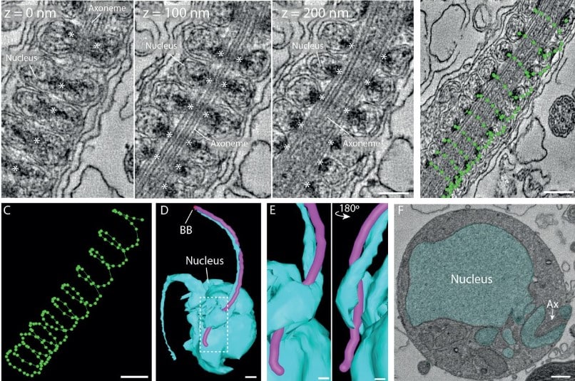
Atypical flagella assembly and haploid genome coiling during male gamete formation in Plasmodium
Molly Hair, Flavia Moreira-Leite, David Ferguson, Mohammed Zeeshan, Rita Tewari, Sue Vaughan
The ultrastructural nature of human oocytes’ cytoplasmatic abnormalities and the role of cytoskeleton dysfunction
Martina Tatíčková, Zuzana Trebichalská, Drahomíra Kyjovská, Pavel Otevřel, Soňa Kloudová, Zuzana Holubcov
Structural evidence for elastic tethers connecting separating chromosomes in crane-fly spermatocytes
Arthur Forer, Shotaro Otsuka
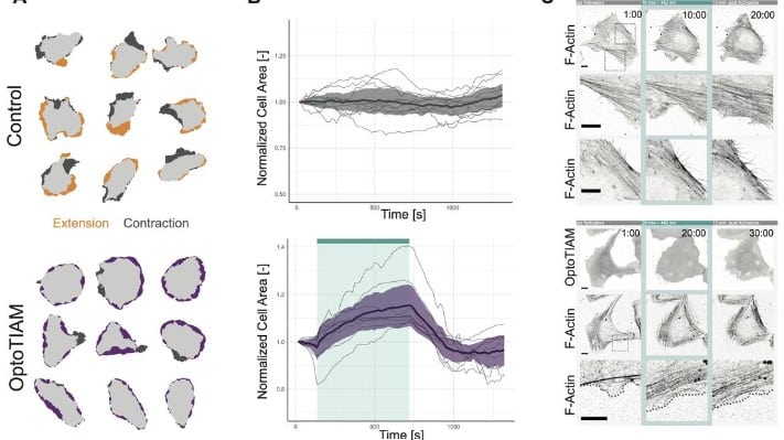
Opto-RhoGEFs: an optimized optogenetic toolbox to reversibly control Rho GTPase activity on a global to subcellular scale, enabling precise control over vascular endothelial barrier strength
Eike K. Mahlandt, Sebastián Palacios Martínez, Janine J. G. Arts, Simon Tol, Jaap D. van Buul, Joachim Goedhart
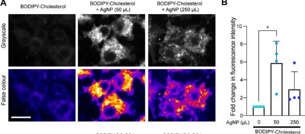
Improved imaging and preservation of lysosome dynamics using silver nanoparticle-enhanced fluorescence
Sumaiya A. Soha, Araniy Santhireswaran, Saaimatul Huq, Jayde Casimir-Powell, Nicala Jenkins, Gregory K. Hodgson, Michael Sugiyama, Costin N. Antonescu, Stefania Impellizzeri, Roberto J. Botelho
Post-transcriptional splicing can occur in a slow-moving zone around the gene
Allison Coté, Aoife O’Farrell, Ian Dardani, Margaret Dunagin, Chris Coté, Yihan Wan, Sareh Bayatpour, Heather L. Drexler, Katherine A. Alexander, Fei Chen, Asmamaw T. Wassie, Rohan Patel, Kenneth Pham, Edward S. Boyden, Shelley Berger, Jennifer Phillips-Cremins, L. Stirling Churchman, Arjun Raj
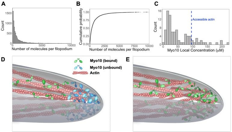
Pushed to the edge: hundreds of myosin 10s pack into filopodia and could cause traffic jams on actin
Julia Shangguan, Ronald S Rock
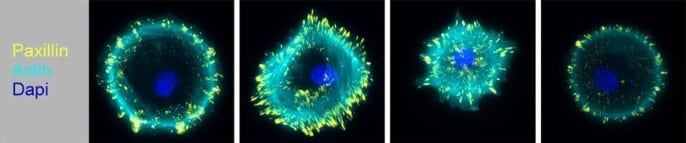
Assessing the performance of the Cell Painting assay across different imaging systems
Callum Tromans-Coia, Nasim Jamali, Hamdah Shafqat Abbasi, Kenneth A. Giuliano, Mai Hagimoto, Kevin Jan, Erika Kaneko, Stefan Letzsch, Alexander Schreiner, Jonathan Z. Sexton, Mahomi Suzuki, O. Joseph Trask, Mitsunari Yamaguchi, Fumiki Yanagawa, Michael Yang, Anne E. Carpenter, Beth A. Cimini
Reorganization of the Flagellum Scaffolding Induces a Sperm Standstill Required for Fertilization
Martina Jabloñski, Guillermina M. Luque, Matías D. Gómez-Elías, Claudia Sanchez-Cardenas, Xinran Xu, Jose Luis de la Vega-Beltran, Gabriel Corkidi, Alejandro Linares, Victor X. Abonza Amaro, Dario Krapf, Diego Krapf, Alberto Darszon, Adan Guerrero, Mariano G. Buffone
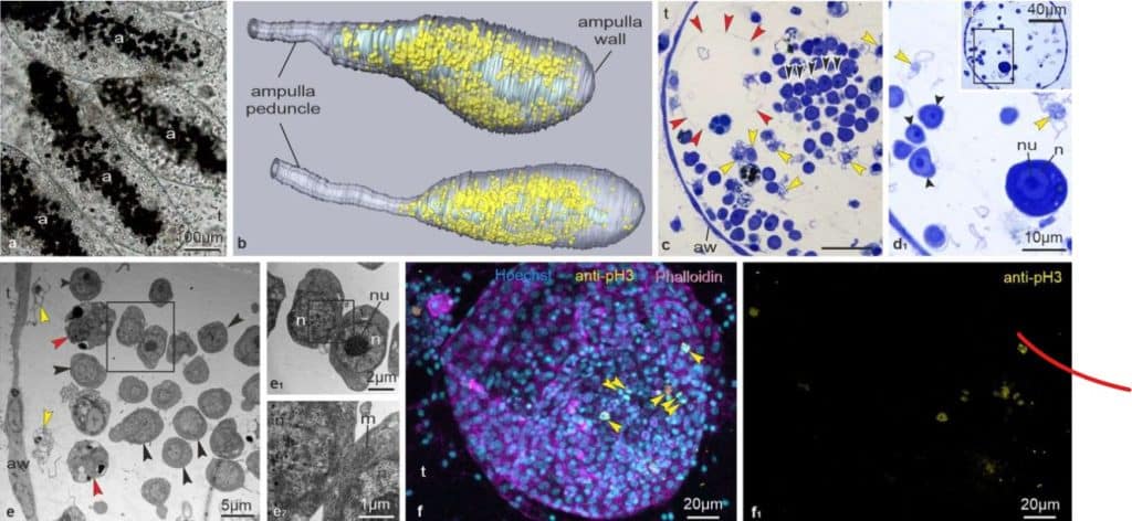
New permanent stem cell niche for development and regeneration in a chordate
Virginia Vanni, Federico Caicci, Anna Peronato, Graziano Martello, Davide Asnicar, Fabio Gasparini, Loriano Ballarin, Lucia Manni
ER-Mitochondria Contact Sites expand during mitosis
Fang Yu, Raphael Courjaret, Asha Elmi, Ayat Hammad, Melanie Fisher, Mark Terasaki, Khaled Machaca
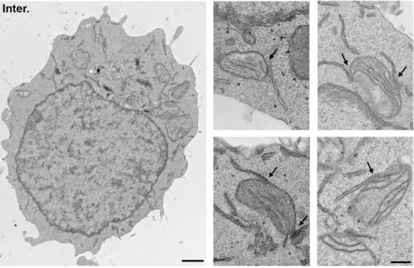
Figure extracted from Yu, et al. . The image is made available under a CC-BY 4.0 International license.
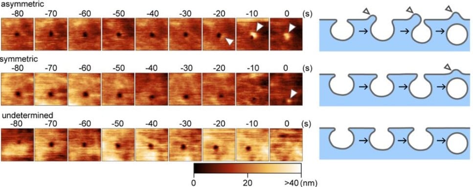
Self-assembly of CIP4 drives actin-mediated asymmetric pit-closing in clathrin-mediated endocytosis
Yiming Yu, Shige H. Yoshimura
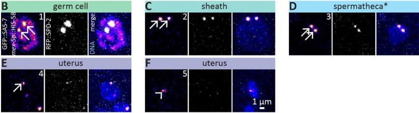
Mapping of centriolar proteins onto the post-embryonic lineage of C. elegans
Nils Kalbfuss, Antonin Berger, Pierre Gönczy
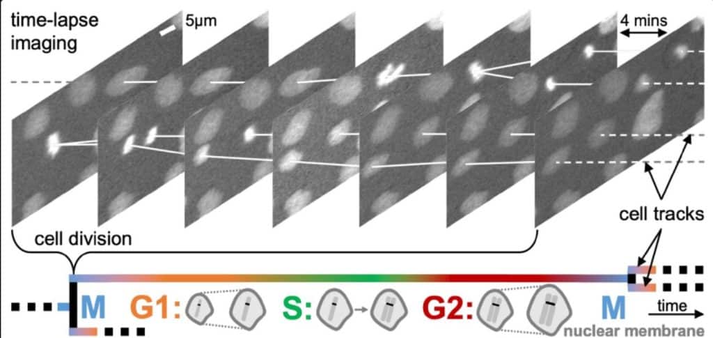
Learning dynamic image representations for self-supervised cell cycle annotation
Kristina Ulicna, Manasi Kelkar, Christopher J Soelistyo, Guillaume T Charras, Alan R Lowe
Glucose-stimulated KIF5B-driven microtubule sliding organizes microtubule networks in pancreatic beta cells
Kai M. Bracey, Pi’illani Noguchi, Courtney Edwards, Alisa Cario, Guoqiang Gu, Irina Kaverina
Single-molecule imaging reveals the kinetics of non-homologous end-joining in living cells
Mariia Mikhova, Joshua R. Heyza, Katheryn Meek, Jens C. Schmidt
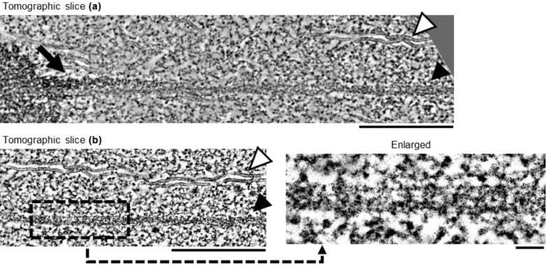
Viscous shear is a key force in Drosophila ventral furrow morphogenesis
Amanda Nicole Goldner, Mohamad Ibrahim Cheikh, Miriam Osterfield, Konstantin Doubrovinski
 (No Ratings Yet)
Get involved
Create an account or log in to post your story on FocalPlane.
More posts like this

Filter by
- NewsApply
- DiscussionsApply
- How toApply
- ToolsApply
- Case studiesApply
- InterviewsApply
- JobsApply
- EducationApply
- Blog seriesApply
- Deep Learning for Bi..o-image analysisApply
- GloBIAS – updates fr..om the communityApply
- WAMBIAN: West Africa.. in FocusApply
- Volume EMApply
- Latin American Micro..scopistsApply
- Bio-image Analysis w..ith NapariApply
- Imaging with…Apply
- Towards Global Acces..sApply
- Latin America Bioima..gingApply
- From Zero to Qupath ..HeroApply
- Asian Microscopists ..and Cell BiologistsApply
- AIC at HHMI JaneliaApply
- Highlights from Euro..-BioImagingApply
- LSFM seriesApply
- DIY MicroscopyApply
- View all