Featured image with Thibault Dhellemmes and Jérémie Teillon
Posted by FocalPlane, on 1 March 2024
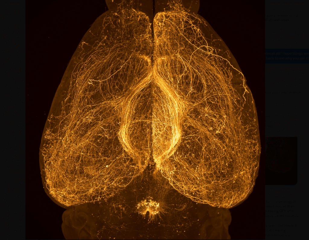
Our featured image shows the full morphology of Relaxin-3 neurons in an entire adult mouse brain. The neurons of interest are expressing GFP after viral injections in a pontine nucleus called nucleus incertus. To observe the full depth of this brain, it was made transparent by applying an optical clearing technique called Adipoclear. This method is a variant of IDISCO+, based on organic solvents. The sample was observed on an Ultramicroscope II light sheet microscope, using a 4x magnification. The image is a maximum intensity projection over the entire depth, created in FIJI.
The sample was prepared by Thibault Dhellemmes and imaged by Jérémie Teillon.
We caught up with Thibault Dhellemmes and Jérémie Teillon to find out more about their research
Thibault Dhellemmes
Current research: Thibault Dhellemmes is a PhD student in the lab of Professor Marc Landry in Neurodegenerative diseases institute (IMN) in Bordeaux. His thesis is about the treatment of chronic pain symptoms and the morphological characterization of the relaxin-3/RXFP3 peptidergic system.
Favourite imaging technique/microscope: Thibault’s favourite technique is the CLEM (correlative light-electron microscopy) approach which consists, in his case, in the association of confocal imaging methods and scanning electron microscopy in array tomography.
What are you most excited about in microscopy? Thibault is interested in high-resolution microscopy techniques. He is impressed by how deep we can get through electron microscopy in the description of neural microcircuits. The nanoscale provided by techniques such as CLEM allow them to picture neurons from the layers of myelin sheets to vesicles and mitochdria.
Jérémie Teillon
Current research: Jérémie Teillon is working as a core facility staff at the Bordeaux imaging Center.He is a specialist in tissue optical clearing and light sheet microscopy of big samples such as whole mouse brain. He supports research projects in neurosciences and cancer research.
Favourite imaging technique/microscope: Jérémie’s favourite methodology is the combination of tissue optical clearing and light sheet microscopy. He is amazed by the impressive imaging depth reachable in that configuration compared to non-transparent samples.
What are you most excited about in microscopy? Jérémie is particularly excited by the image analysis of full mouse brains. Recent developments have shown that it is possible to register the brain 3D images on reference atlases such as the Allen Brain Atlas. This provides a new way to study brain connectivity or cells distribution in an unbiased way. Moreover, it is much faster compared to traditional slice-based histology methods.
 (3 votes, average: 1.00 out of 1)