Featured image with Ciarán Butler Hallissey
Posted by FocalPlane, on 13 September 2024
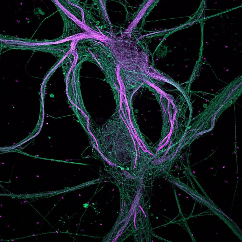
Our featured image, acquired by Ciarán Butler Hallissey, is a maximum-intensity projection of rat hippocampal neurons processed with ultrastructure expansion microscopy (U-ExM) and captured with a spinning disk confocal microscope.
Find out more about the image and Ciarán’s research below.
More about the image: Expansion microscopy physically increases the size of your sample to improve the resolution. It means you can get super-resolution without super-resolution microscopes, however, I’m very fortunate in that I can use both together! The expansion protocol I used is U-ExM which was developed in the Guichard-Hamel Lab at the University of Geneva. U-ExM expands the sample approximately four times in all dimensions and is followed by immunostaining. This means that antibodies have more space to wiggle around and can more densely label the sample therefore improving image quality. In this image, you see Microtubule-associated protein 2 (MAP2) in magenta and microtubules in green. As its name indicates MAP2 interacts with microtubules, but more specifically it interacts with microtubules in the neuron cell body and the dendrites but not in the axon. So, what we see here is a neuron intertwined in a sea of axons!
Research career so far: My career has revolved around visualizing increasingly smaller structures with the use of a wide variety of microscopy techniques. It started with my BSc majoring in Anatomy at the University of Galway, Ireland. This was where I first started my microscopy journey with histology and using confocal microscopy to visualize inducible DNA damage. At the end of my degree, I worked in the same Anatomy Department the following year, teaching anatomy to science and medical students while also working on projects using both fluorescence and electron microscopy. After this, I did an MSc in biomedical imaging at the University of Turku, Finland, again diving deeper into fluorescence microscopy while establishing colon organoid cultures. I then joined the local microscopy facility at Turku Bioscience as a research engineer, training people and maintaining a wide array of fluorescence microscopes. I learned a lot more about microscopy technologies while also supporting many research projects from a very different angle that I had not experienced before. Currently, I’m doing a PhD in the NeuroCyto Lab at Aix-Marseille University, France.
Current research: I’m studying the microtubules inside hippocampal neurons. More specifically, I’m looking at the roles that microtubules have in developing neurons and how they are maintained throughout their life. Within this development and maintenance, I’m looking at how the microtubules are repaired and renewed. We know a lot about how microtubules can grow and shrink at their ends but the topic of repair along the microtubule has only recently been seen within cells. We have a recent publication on this here. I’m visualizing this microtubule repair inside neurons that are incredibly small and packed with microtubules. The aim is to look inside the neurons and see microtubule repair at a single microtubule level. This is why expansion microscopy and other super-resolution methods are crucial for me.

Favourite imaging technique/microscope: My favourite microscope is where the eyepieces and the desk height for the monitor are at the same level, so I don’t need to increase the height of the chair or half stand to use the eyepieces. But in general, I like many techniques. I am usually most interested in the technique that helps me answer my question. So right now, it’s expansion microscopy which takes more sample preparation but has great benefits for my project. I do have a soft spot for light-sheet because of the flexibility, imaging speed and gentle illumination on live cells. But we need to find more ways of handling the massive data that it generates.
What are you most excited about in microscopy? From instrumentation to image analysis, the process of capturing and analysing images involves a diversity of researchers from physicists, biologists, computer scientists and more. We have an ever-growing abundance of specialized imaging and analysis approaches but getting them into the hands of everyday research is challenging. Fortunately, we have a growing community putting a huge effort into translating this specialized knowledge into a common language and connecting everyone for discussion. This attracts a wider audience which then improves knowledge transfer and feedback cycle for future improvements. I’m excited to see more of this and see the terms like ‘advanced’ removed from many approaches.
 (No Ratings Yet)