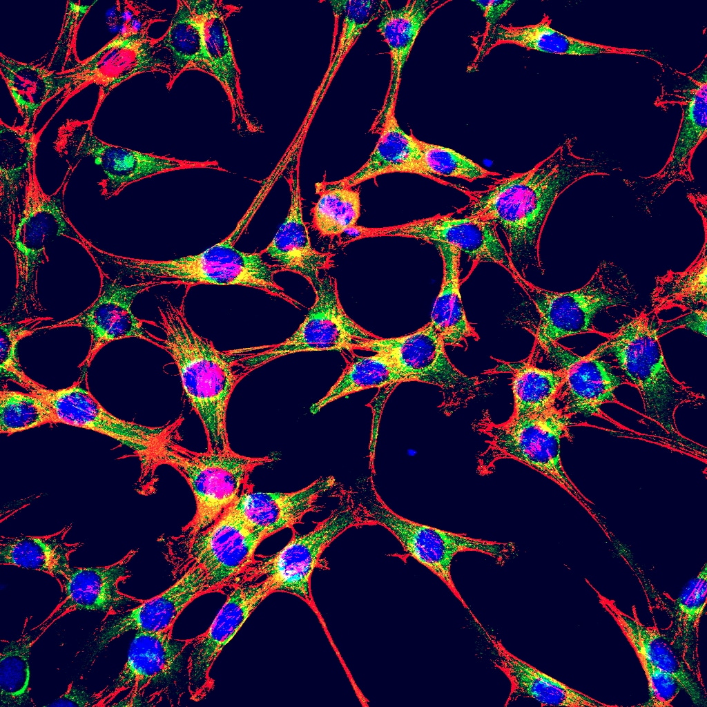
Featured image with Jyoti Maddhesiya
Posted by FocalPlane, on 8 November 2024
Our featured image, acquired by Jyoti Maddhesiya, is a laser scanning confocal image of H9c2 cardiomyoblast cells. The green shows BMP7 in the cytoplasm, the blue highlights the nuclei and the cytoskeleton protein F-actin is labelled in red. The cells were transfected with a BMP7 construct and fixed and labelled 48 hours post transfection. The cells were imaged using a Leica SP8 STED microscope at the Central Discovery Centre, Banaras Hindu University. The image was analysed using ImageJ and presented as maximum intensity projection.

Find out more about Jyoti’s research below:
Research career so far: In childhood, I loved drawing and capturing colourful images. From the very beginning of my studies I was interested in science, especially zoology. I graduated with an undergraduate degree in biology and completed my master’s in zoology at Deen Dayal Upadhyay University, Gorakhpur. During my degree, I was curious to understand how different diseases are caused and the possible mechanism and factors contributing to the disease. My undergraduate project was on fasciolosis disease, one of the common diseases caused by liver flukes. Through this project I became enthusiastic to continue doing research. In my masters, I was keen to continue with this type of research but, unfortunately, I was allocated a project on exotic fishes. I was excited to start my PhD where my project uses clinical, epidemiological, and molecular techniques. When I started working in a cytogenetics lab, I was intrigued by the research on Drosophila models and scanning their different features using confocal microscopy. This introduced me to using confocal microscopy in my research.
Current research: Currently, I am pursuing PhD on Cardiovascular Genetics under Prof. Bhagyalaxmi Mohapatra in Banaras Hindu University (Varanasi, India). My current project title is ‘Genetic and non-genetic factors involved in congenital heart disease.’ The core part of the project is based on the mutational screening of selected cardiac genes identified in congenital heart disease patients. I use different assays including in-silico, biochemical, gene and protein expression analysis to examine these mutations, and other novel mutations that we have identified. We have used confocal microscopy to examine the localization of wild type BMP7 and we compare this with the expression and localization of mutated proteins.
Favourite imaging technique/microscope: I like the fixation technique used to label proteins. Through this technique, different features of cells can be stained with vibrant colours. My favourite microscope is the Leica SP8 STED Laser Scanning Super Resolution confocal microscope. I have also used Zeiss LSM 780 but because the Leica SP8 is an inverted microscope, it is user-friendly and it has advance properties with higher magnification and live cell imaging feature.
What are you most excited about in microscopy? This image of BMP7 expressing cells is the most electrifying bioimaging that allowed me to visualise the details of our basic unit – the cell – using the confocal microscopy. The higher magnification of Leica SP8 allow us to visualize in depth the colocalization of BMP7 protein with ER-tracker. This was thrilling to us as it has not been reported previously. However, I am looking forward for the forthcoming advancements in confocal microscopy that allow us to depict the detailing of organelles more precisely.


 (No Ratings Yet)