Featured image with Omkar Joshi
Posted by FocalPlane, on 14 March 2025
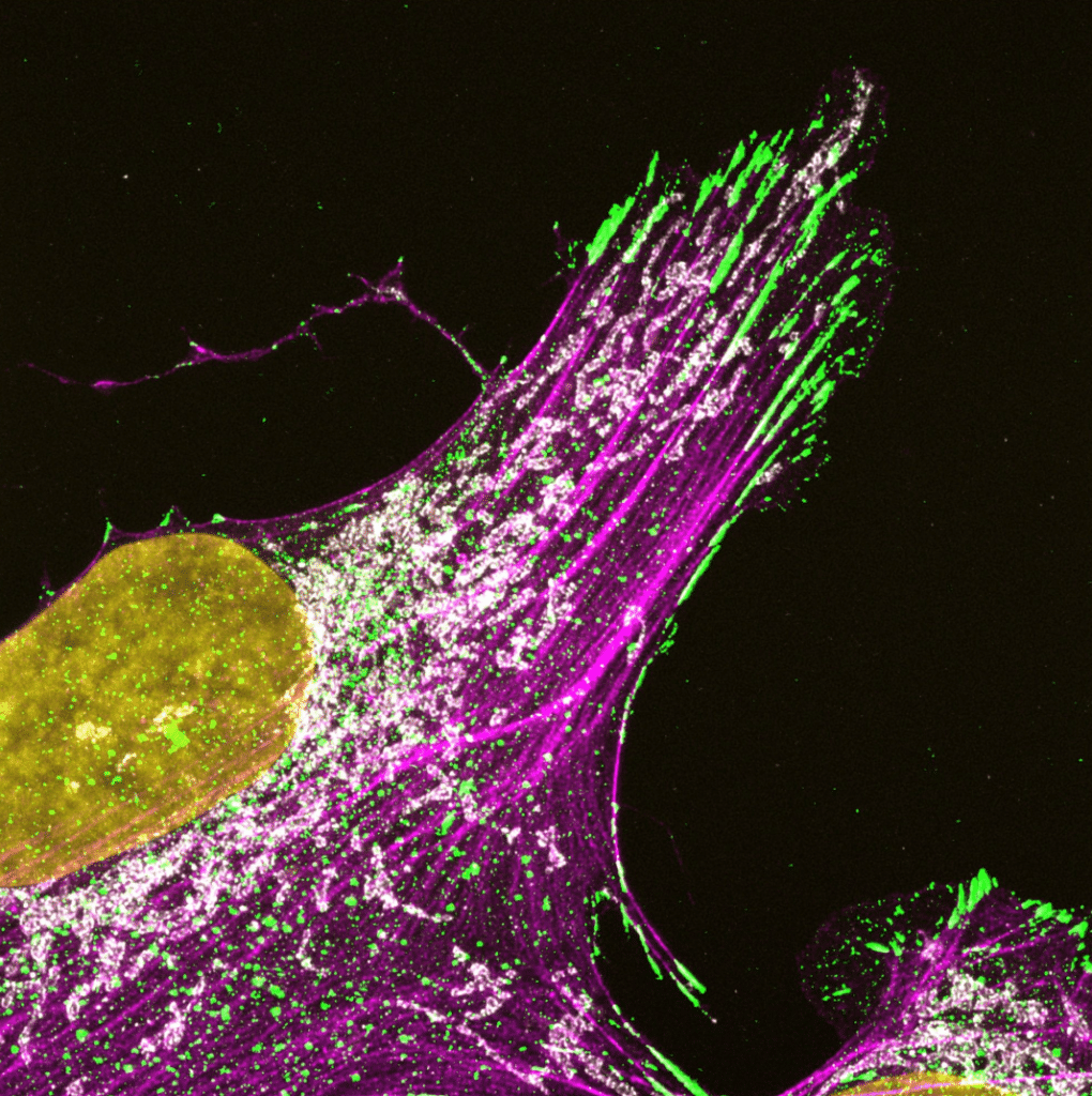
Our featured image, acquired by Omkar Joshi, showcases a U2OS cell immunostained for paxillin (green), TOMM20 (mitochondria, white), and F-actin (labelled using SiR-Actin, magenta), with DAPI (yellow) marking the nucleus. The sample was fixed with 4% PFA, followed by immunostaining with appropriate primary and secondary antibodies. Imaging was performed using a Zeiss LSM880 with AiryScan, and the final image was deconvolved using Huygens Professional software.
Discover more about Omkar’s research
Research career so far: I am currently a PhD researcher working in the lab of Professor Johanna Ivaska at the Turku Bioscience Center of the University of Turku, Finland. Before this, I completed a Bachelor of Science-Master of Science (BS-MS) Dual Degree at the Indian Institute of Science Education and Research (IISER), Pune. I am a cell biologist by training, and I have always been curious by how different signaling pathways regulate cellular behaviors—an interest that has carried through from my undergraduate studies to my PhD research.
Current research: My current PhD research focuses on the underappreciated crosstalk between cell-ECM adhesion and mitochondria. Using a combination of quantitative microscopy, biochemical techniques and cell biological assays, I investigate how this interplay impacts processes like focal adhesion dynamics, cell spreading and cancer cell migration.
Favourite imaging technique/microscope: It’s hard to pick just one! I’ve had the opportunity to use a few different imaging systems, especially here in Turku. It is, of course, awesome to get pretty pictures using super-resolution techniques like SIM or AiryScan, but others such as FLIM can reveal more intricate phenomena. I am interested in several of the existing imaging systems, especially SMLM systems, and look forward to exploring them in the future!
What are you most excited about in microscopy? The growing integration AI in microscopy is very exciting. This pertains not only to analysis pipelines, but also capturing images in real time. Bridging the gap between throughput and resolution by further developing adaptive microscopy approaches promises to be a very interesting avenue currently and in the future.
 (8 votes, average: 1.00 out of 1)
Interesting topic. Has potential to change the course of cancer prognosis