Microscopy preprints – new tools and techniques in imaging
Posted by FocalPlane, on 2 May 2025
Here is a curated selection of preprints posted recently on new tools and techniques in imaging. Let us know if we are missing any recent preprints that are on your reading list!
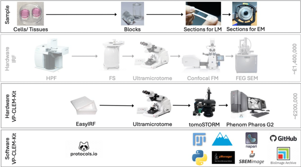
VP-CLEM-Kit: An accessible pipeline for visual proteomics using super resolution volume correlative light and electron microscopy (SR-vCLEM)
Dumisile Lumkwana, Jonathan Lightley, Arturo G. Vesga, Joost de Folter, Marie-Charlotte Domart, Catherine Maclachlan, Sunil Kumar, James Evans, Christopher Peddie, Alana Burrell, Azumi Yoshimura, Asandile Mangali, Nicola Vahrjmeijer, Meenal Bhaga, Catherine Smit, Benjamin Titze, Mathew Horrocks, Lorian Cobra Straker, Edwin Garcia, Martin Jones, Adam Mclean, Candice Roufosse, Ben Loos, Sonia Gandhi, Amy Strange, Ricardo Henriques, Paul French, Lucy Collinson
Effects of base temperature, immersion medium, and EM grid material on devitrification thresholds in cryogenic optical super-resolution microscopy
Soheil Mojiri, Joseph M. Dobbs, Niko Faul, Thomas P. Burg, Julia Mahamid, Jonas Ries
FilaBuster: A Strategy for Rapid, Specific, and Spatiotemporally Controlled Intermediate Filament Disassembly
Andrew S. Moore, Tommy Krug, Simon B. Hansen, Alexander V. Ludlow, Jonathan B. Grimm, Anthony X. Ayala, Sarah E. Plutkis, Nan Wang, Robert D. Goldman, Ohad Medalia, Luke D. Lavis, David A. Weitz, Jennifer Lippincott-Schwartz
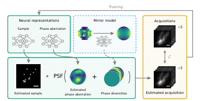
DeepPD: Joint Phase and Object Estimation from Phase Diversity with Neural Calibration of a Deformable Mirror
Magdalena C. Schneider, Courtney Johnson, Cedric Allier, Larissa Heinrich, Diane Adjavon, Joren Husic, Patrick La Rivière, Stephan Saalfeld, Hari Shroff
Improving single molecule localisation microscopy reconstruction by extending the temporal context
Sebastian Reinhard, Vincent Ebert, Jann Schrama, Markus Sauer, Philip Kollmannsberger
Quantifying Uncertainty in Phasor-Based Time-Domain Fluorescence Lifetime Imaging Microscopy
Qinyi Chen, Jongchan Park, Shuqi Mu, Liang Gao
Combining quantum cascade lasers and plasmonic metasurfaces to monitor de novo lipogenesis with vibrational contrast microscopy
Steven H. Huang, Dias Tulegenov, Gennady Shvets
Three-dimensional Optical Reconstruction of colloidal electrokinetics via multiplane imaging
Flip de Jong, Pablo Diez-Silva, Jui-Kai Chen, Raúl Pérez-Peláez, Sudipta Seth, Harishankar Balakrishnan, Bing-Yang Shih, Maarten Rosmeulen, Santi Nonell, Susana Rocha, Andrey Klymchenko, Luis Liz-Marzán, Roger Bresolí-Obach, Manuel I. Marqués, Rafael Delgado Buscalioni, Johan Hofkens, Boris Louis
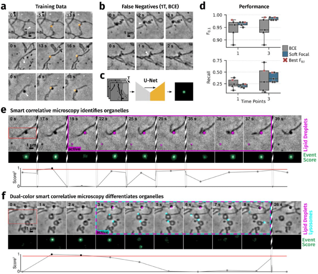
Smart hybrid microscopy for cell-friendly detection of rare events
Willi L. Stepp, Emine Berna Durmus, Santiago N. Rodriguez Alvarez, Juan C. Landoni, Giorgio Tortarolo, Kyle M. Douglass, Martin Weigert, Suliana Manley
Microbubble Backscattering Intensity Improves the Sensitivity of Three-dimensional (3D) Functional Ultrasound Localization Microscopy (fULM)
Qi You, YiRang Shin, Yike Wang, Matthew R. Lowerison, Bing-Ze Lin, Pengfei Song
Expansion-assisted selective plane illumination microscopy for nanoscale imaging of centimeter-scale tissues
Adam Glaser, Jayaram Chandrashekar, Sonya Vasquez, Cameron Arshadi, Rajvi Javeri, Naveen Ouellette, Xiaoyun Jiang, Judith Baka, Gabor Kovacs, Micah Woodard, Sharmishtaa Seshamani, Kevin Cao, Nathan Clack, Andrew Recknagel, Anna Grim, Pooja Balaram, Emily Turschak, Marcus Hooper, Alan Liddell, John Rohde, Ayana Hellevik, Kevin Takasaki, Lindsey Erion Barner, Molly Logsdon, Chris Chronopoulos, Saskia de Vries, Jonathan Ting, Steve Perlmutter, Brian Kalmbach, Nikolai Dembrow, Bosiljka Tasic, R. Clay Reid, David Feng, Karel Svoboda
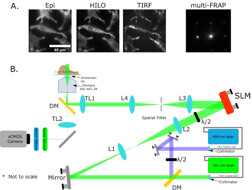
Combining Multi-site FRAP and HILO-TIRF microscopy using a Spatial Light Modulator
Avinash Upadhya, Yean Jin Lim, Woei Ming Lee
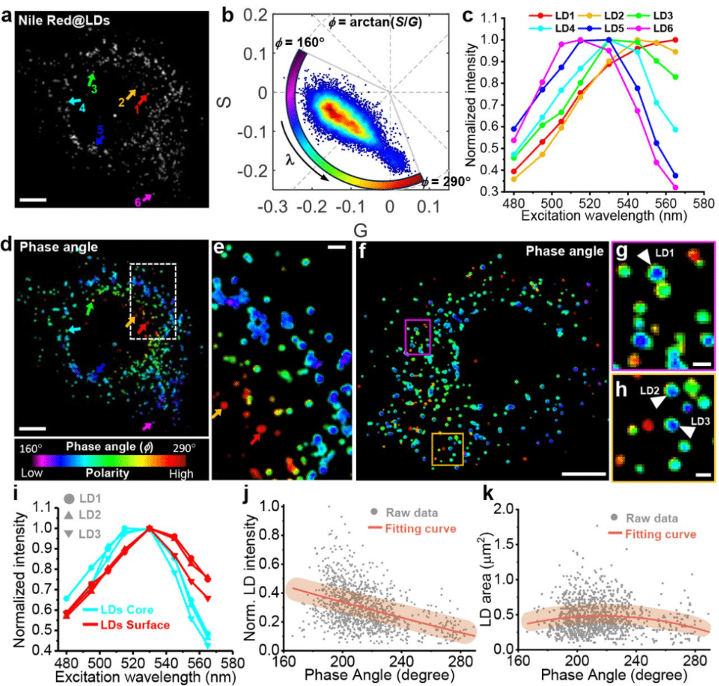
Excitation Spectral Phasor Microscopy (ExSPM) unveils spatiotemporal heterogeneities in polarity of intracellular lipid droplets
Jiayi Liu, Yi He, Rui Yan, Jinhong Yan, Ruirong Wang, Zhipeng Zhang, Jiankai Xia, Igor Kireev, Ke Xu, Kun Chen
Optical sectioning for reflection interference microscopy: quantitative imaging at soft interfaces
Cathie Ventalon, Oksana Kirichuk, Yotam Navon, Yan Chastagnier, Laurent Heux, Ralf P. Richter, Lionel Bureau, Delphine Débarre
Super-resolving particle diffusion heterogeneity in porous hydrogels via high-speed 3D active-feedback single-particle tracking microscopy
Yuxin Lin, Haoting Lin, Kevin Welsher
Altair-LSFM: A High-Resolution, Easy-to-Build Light-Sheet Microscope for Sub-Cellular Imaging
John Haug, Seweryn Gałecki, Kevin M. Dean
Ab-trapping – a peripheral staining artifact in antibody-based microscopy and genomics
Konrad Chudzik, Yuko Sato, Xingchi Yan, Simon Ullrich, Watanya Trakarnphornsombat, Lothar Schermelleh, Geoffrey Fudenberg, Hiroshi Kimura, Michael I. Robson, Irina Solovei
A genetic strategy to allow detection of F-actin by phalloidin staining in diverse fungi
Alison CE Wirshing, Analeigha V Colarusso, Daniel J Lew
Mesoscopic SCAPE Microscope with a Rescanned, Super-oblique Illumination Plane
Zixian Cao, Zhu Jiapeng, Cheng Zhang, Qianqian Wang, Yankan Huang, Wei Liu, Bingxin Shen, Yuming Chai, Zhaoming Zhong, He Li, Quan Wen, Han Wang, Wenxuan Liang
Deep-Tissue Two-Photon Brain Imaging Enabled by a Tunable Fiber-Optic Dispersive Wave Generator
Marvin Edelmann, Andreu Matamoros-Angles, Mohsin Shafiq, Mikhail Pergament, Markus Glatzel, Franz X. Kärtner
Nickel-NTA lipid-monolayer affinity grids allow for high-resolution structure determination by cryo-EM
Aleksandra Skrajna, Emily Robinson, Kevin Cannon, Reta Sarsam, Richard G. Ouellette, Patrick Brennwald, Robert K. McGinty, Joshua D. Strauss, Richard W. Baker
Mind the corner: Fillets in cryo-FIB lamella preparation to minimise sample loss caused by stress concentration and lamella breakage
Sergey Gorelick, Sailakshmi Velamoor, Patrick Cleeve, Sylvain Trépout, Le Ying, Vivek Naranbhai, Georg Ramm
Exploring shaped focused ion beams for lamella preparation
Johann Brenner, Jürgen M. Plitzko, Sven Klumpe
A Versatile and Open Source One- and Two-Photon Light-Sheet Microscope Design
Antoine Hubert, Thomas Panier, Geoffrey Migault, Hugo Trentesaux, Benoît Beaudou, Georges Debrégeas, Volker Bormuth
A non-toxic, user-friendly buffer that enhances green fluorophore performance in conventional and super-resolution imaging
Anaïs C. Bourges, Robin Van den Eynde, Wim Vandenberg
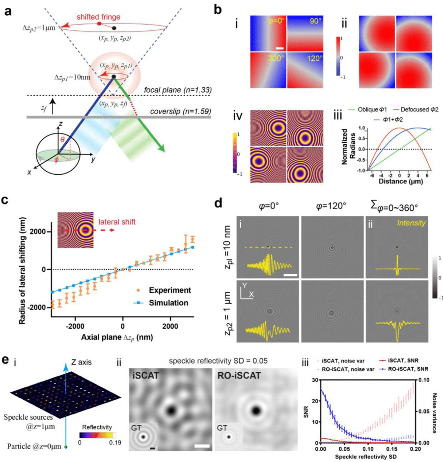
Using rotational integration of oblique interferometric scattering (RO-iSCAT) to track axial spatiotemporal responses of membrane protrusions
Junyu Liu, Yean Jin Lim, David Herrmann, Paul Timpson, Tri G. Phan, Huafeng Liu, Min Guo, Woei Ming Lee
A Beauty Dye Staining (ABDS) – inexpensive marker-ink based open-source alternative for commercial membrane vital dyes
Anna A. Abelit, Natalia A. Boitsova, Liudmila E. Yakovleva, Anton A. Kornev, Daniil D. Stupin
Imaging throughput of compact handheld microscopes for quantitative single cell studies
Sophie Bulloch, Tienan Xu, David Herrmann, Paul Timpson, Tri Giang Phan, Yean Jin Lim, Woei Ming Lee
Evaluating MINFLUX experimental performance in silico
Zach Marin, Jonas Ries
SpeedyTrack: Direct microsecond wide-field single-molecule tracking and super-resolution mapping via CCD vertical shift
Megan A. Steves, Ke Xu
Transient optical clearing using absorbing molecules for ex-vivo and in-vivo imaging
Muhammed Waqas Shabbir, Matthew Phillips, David Asante-Asare, Zihao Ou
Aberration correction in long GRIN lens-based microendoscopes for extended field-of-view two-photon imaging in deep brain regions
Andrea Sattin, Chiara Nardin, Simon Daste, Monica Moroni, Innem Reddy, Carlo Liberale, Stefano Panzeri, Alexander Fleischmann, Tommaso Fellin
A modular multi-color fluorescence microscope for simultaneous tracking of cellular activity and behavior
Euphrasie Ramahefarivo, Leonard Böger, Takkasila Saichol, Behzad Shomali, Luis Alvarez, Monika Scholz
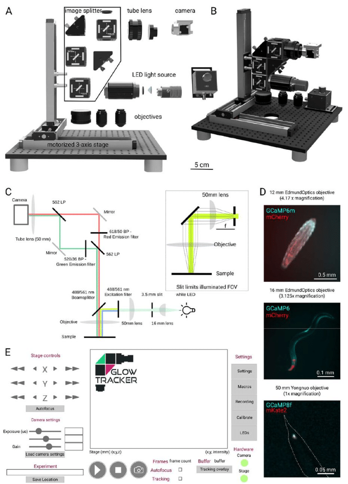
A novel epifluorescence microscope design and software package to record naturalistic behaviour and cell activity in freely moving Caenorhabditis elegans
Sebastian N. Wittekindt, Hannah Owens, Lennard Wittekindt, Aurélie Guisnet, Michael Hendricks
Optimizing Tissue Clearing Methods for Improved Imaging of Whole-Mount Retinas
Aubin Mutschler, Volha V. Malechka, Petr Baranov, Jonathan R. Soucy
Real-Time Wide-Field Fluorescence Lifetime Imaging via Single-Snapshot Acquisition for Biomedical Applications
Vikas Pandey, Euan Millar, Ismail Erbas, Luis Chavez, Jack Radford, Isaiah Crosbourne, Mansa Madhusudan, Gregor G. Taylor, Nanxue Yuan, Claudio Bruschini, Stefan T. Radev, Margarida Barroso, Andrew Tobin, Xavier Michalet, Edoardo Charbon, Daniele Faccio, Xavier Intes
 (No Ratings Yet)