Featured image with Andrew Octavian Sasmita
Posted by FocalPlane, on 23 May 2025
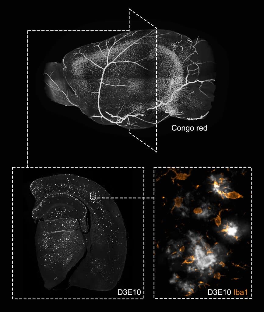
Our featured image, acquired by Andrew Octavian Sasmita, depicts a single brain hemisphere of the 5xFAD mouse model of Alzheimer’s disease (AD) at different spatial scales. At the whole tissue and 3D level, the unequal distribution of Congo red-labelled amyloid-β (Aβ) plaque load is evident across different brain regions and throughout the brain’s axes (top image). A closer 2D inspection reveals similar regional differences in plaque distribution, labelled with the D3E10 Aβ antibody (bottom left image), due to distinct transgene activity in different neuronal clusters. Visualisation of single Aβ plaques and co-labelling with markers for other cell types (e.g., microglia, Iba1) further highlights the complex and dense environment shared between extracellular Aβ plaques and neural cells, as well as their crosstalk (bottom right image).
Preparation and imaging: After organ collection from a 6-month-old female animal, the brain was processed for either light-sheet imaging or epifluorescence imaging. The top image is a 3D render of the Congo red-labelled and optically cleared brain (using a modified iDISCO protocol) obtained from tiled and serial Z-stack imaging with the UltraMicroscope II light-sheet system. The bottom images were captured using the Zeiss Observer Z1 microscope equipped with a Plan-Apochromat ×20/0.8 objective and the Colibri LED system. The bottom left image is a tiled image of an entire coronal section of the paraffin-embedded hemibrain, while the bottom right image is a zoomed-in view of Aβ plaques and corralling microglia.
Discover more about Andrew’s research
Research career so far: I completed my PhD in the Department of Neurogenetics at the Max Planck Institute for Multidisciplinary Sciences, where I primarily investigated whether oligodendrocytes, the myelinating glia of the central nervous system, produce Aβ and contribute to plaque formation in AD models. During this time, I also explored the intricate relationship between neurons and glia in health and disease, as well as the fundamental research gaps in the basic pathophysiology of AD. I am currently a postdoctoral researcher in the Department of Anatomy and Neuroscience at University College Cork.
Current research: I am now studying the potential contribution of the gut microbiome in mediating sex-specific differences in AD pathology, with a focus on glial responses and adult hippocampal neurogenesis.
Favourite imaging technique/microscope: It’s a tie between the Zeiss Observer system and light-sheet microscopy.
What are you most excited about in microscopy? Multimodal, high-resolution, and high-throughput systems have always been near and dear to me, especially in quantitative research. I am also excited about future developments in small fluorescent protein tags, particularly in combination with super-resolution microscopy, to better understand the localisation of proteins within compact cellular compartments.
 (No Ratings Yet)