Workshop report: UC Berkeley’s Advanced Imaging Methods Workshop
Posted by Ben Cox, on 29 May 2025
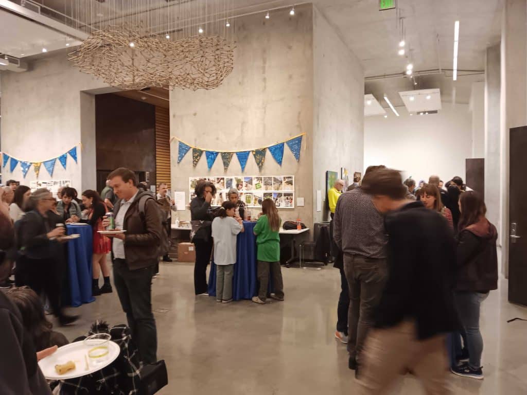
I attended UC Berkeley’s Advanced Imaging Methods Workshop with goals of becoming more familiar with a) high resolution imaging methods and hardware I did not have access to at my home institution and b) image processing and analysis algorithms, especially those based on machine learning/AI. The workshop provided ample opportunity to fulfill both of these goals.
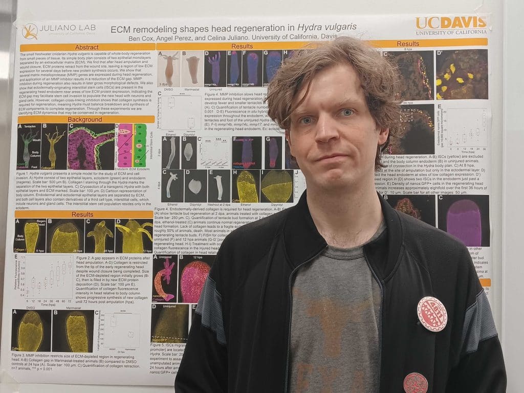
Of the many types of imaging hardware described and demonstrated at the workshop, I was particularly interested in two- and three-photon imaging. I study extracellular matrix (ECM) in an organism (Hydra vulgaris) that has fewer transgenic tools than more established models, so I am intrigued by the possibility of using these microscopes for second and third harmonic generation imaging of the ECM protein collagen. Marco Arrigoni from Light Conversion and Dan Giardina of 3i gave compelling presentations on lasers sources optimized for two- and three-photon imaging, and I attended a demonstration of a Bessel Two-Photon system where I was able to see neurons in a wider z plane than available through conventional Gaussian focus. As I hope to live image collagen remodeling in 3D, learning of this technique gave me greater confidence in the feasibility of some of my planned experiments. Further, the Tomocube HT-X1 demo at the workshop gave me ideas for additional experiments. While my conversations with the representatives there indicated its diffraction-based imaging may not be ideal for studying ECM, I also want to use live imaging to capture migration and invasion of cells through epithelial layers, and this low-toxicity imaging that has the benefit of not requiring fluorescent transgenic animals could facilitate such experiments.
While new hardware could open up new avenues of research for me, I have managed to accumulate considerable fluorescent imaging data from my institution’s confocal core. Thus, my second goal in attending AIM 2025 was to learn about newer image segmentation and analysis algorithms. The second day of the workshop was particularly relevant to my interests, with representatives from microscope companies (ZEISS, Leica, Oxford Instruments, etc.) providing overviews of their proprietary software. Further, I saw presentations from scientists from labs at research institutions have made their image analysis code, itself built on open-source software like cellpose, publicly available via GitHub. Based on these presentations, I hope to test several of these software options, particularly Chan Zuckerberg Biohub’s Omega and Zeiss’s Airivis Pro, on image stacks I have already acquired with the hope that they can be trained to segment cells with greater success than the software I have used in the past.
In summary, my attendance at AIM 2025 was a success on both fronts: it provided ideas for how new hardware could enable future experiments—especially once I have started my own lab and have a budget for microscopes—and how open-source and commercially available software could streamline analysis of imaging data I already have.
JCS-FocalPlane Training Grants are open to all early-career scientists working in the field of cell biology. They can be used to attend training courses in microscopy and bioimage analysis. Drop us an email at focalplane@biologists.com if you have any questions about the grants.
 (No Ratings Yet)
Get involved
Create an account or log in to post your story on FocalPlane.
More posts like this

Filter by
- NewsApply
- DiscussionsApply
- How toApply
- ToolsApply
- Case studiesApply
- InterviewsApply
- JobsApply
- EducationApply
- Blog seriesApply
- Bio-image Analysis w..ith NapariApply
- Imaging with…Apply
- Towards Global Acces..sApply
- Latin America Bioima..gingApply
- From Zero to Qupath ..HeroApply
- Asian Microscopists ..and Cell BiologistsApply
- AIC at HHMI JaneliaApply
- Deep Learning for Bi..o-image analysisApply
- GloBIAS – updates fr..om the communityApply
- Volume EMApply
- Latin American Micro..scopistsApply
- Highlights from Euro..-BioImagingApply
- LSFM seriesApply
- DIY MicroscopyApply
- View all