Microscopy preprints – new tools and techniques in imaging
Posted by FocalPlane, on 25 July 2025
Here is a curated selection of preprints posted recently on new tools and techniques in imaging. Let us know if we are missing any recent preprints that are on your reading list!
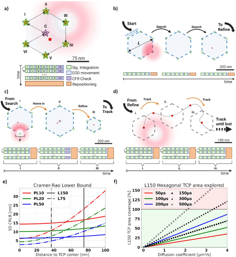
Parameter Optimization for Iterative MINFLUX Microscopy enabled Single Particle Tracking
Bela T.L. Vogler, Giovanni De Angelis, Ziliang Zhao, Christian Eggeling, Francesco Reina
Pulsed-laser lensing for phase modulation in electron microscopy
Daniel X. Du, Adam C. Bartnik, Cameron J. R. Duncan, Usama Choudhry, Tanya Tabachnik, Chaim Sallah, Yuki Ogawa, Ebrahim Najafi, Ding-Shyue Yang, Jared M. Maxson, Anthony W. P. Fitzpatrick
Blood Vessel-Inspired Surface-Emitting Microscopy for Label-Free Monitoring of Single Cell Dynamics
Yu-Cheng Chen, Chaoyang Gong, Guocheng Fang, Jun Xie, Guang Yang, Song Zhu, Zhen Qiao,Yuan Gong
Imaging of specialized plant cell walls by improved cryo-CLEM and cryo-electron tomography
J Daraspe, E Bellani, D De Bellis, C Genoud, N Geldner
Longitudinal Awake Imaging of Mouse Deep Brain Microvasculature with Super-resolution Ultrasound Localization Microscopy
Yike Wang, Matthew R. Lowerison, Zhe Huang, Qi You, Bing-Ze Lin, Daniel A. Llano, Pengfei Song
Non-Invasive Mechanical-Functional Analysis of Individual Liver Mitochondria by Atomic Force Microscopy
Ekaterina O. Zorikova, Sabita Chourasia, Irit Rosenhek-Goldian, Sidney R. Cohen, Semen V. Nesterov, Atan Gross
Rapid and Efficient Quality Control Analysis of Isolated Mitochondria by Interferometric Light Microscopy
Sabah Mozafari, Christopher Ribes, Dmitry Ayollo, Florence Gazeau, Amanda K. A. Silva, Kelly Aubertin
High-speed whole-brain imaging in Drosophila
Wayan Gauthey, Albert Lin, Osama M. Ahmed, Andrew M. Leifer, Mala Murthy, Stephan Y. Thiberge
MitoTracker transfers from astrocytes to neurons independently of mitochondria
Katriona L. Hole, Rosalind Norkett, Emma Russell, Molly Strom, Jack H. Howden, Nicola J. Corbett, Janet Brownlees, Michael J. Devine
De novo designed bright, hyperstable rhodamine binders for fluorescence microscopy
Yuda Chen, Klaus Yserentant, Kibeom Hong, Yiming Kuang, Arghya Bhowmick, Arthur Charles-Orszag, Samuel J. Lord, Lei Lu, Kaipeng Hou, Samuel I. Mann, Jonathan B. Grimm, Luke D. Lavis, R. Dyche Mullins, William F. DeGrado, Bo Huang
A lentiviral fluorescent reporter to study circadian rhythms in single cells
Christian H. Gabriel, Luis Lehmann, Joana Ahlburg, Achim Kramer
CALIPERS: Cell cycle-aware live imaging for phenotyping experiments and regeneration studies
Moises Di Sante, Melissa Pezzotti, Julius Zimmermann, Alessandro Enrico, Joran Deschamps, Elisa Balmas, Silvia Becca, Samantha Solito, Alessandro Reali, Alessandro Bertero, Florian Jug, Francesco S. Pasqualini
Beyond Static Screens: A High-Throughput Pooled Imaging CRISPR Platform for Dynamic Phenotype Discovery
Sravasti Mukherjee, Menno van Tooren, Giulia Zanetti, Dimitris Sfakianakis, Dominique Kemps, Jeffrey Klarenbeek, Hendrik J. Kuiken, Cor Lieftink, Bram van den Broek, Roderick L. Beijerbergen, Kees Jalink
mScarlet3-H with low brightness and fluorescence lifetime has potential for cellular lifetime-unmixing and lifetime-based pH-sensing applications
Theodorus W.J. Gadella, Laura van Weeren
Fast, Bright and Reversible Rhodamine Tags for Live-Cell Imaging
Julian Kompa, Lars Jeremy Dornfeld, Nicola Porzberg, Soohyen Jang, Simon Hans Lilje, Claudia Catapano, David Jocher, Lukas Merk, Silja Zedlitz, Runyu Mao, Jonas Wilhelm, Marina Dietz, Miroslaw Tarnawski, Julien Hiblot, Mike Heilemann, Kai Johnsson
Multiparametric Correlative Topographical and Volumetric Fluorescence Microscopy
Wenzhi Hong, Ziwei Zhang, Ao Li, Ting Sun, Yunzhao Wu, Devkee M. Vadukul, Dylan Jones, Bing Li, Fengjie Liu, Francesco A. Aprile, Julia Gorelik, David Klenerman, Andrew Shevchuk
Versatile Vasculature Chips for Ultrasound Localization Microscopy
Renxian Wang, Qi Liu, Xin Zhao, Wei-Ning Lee
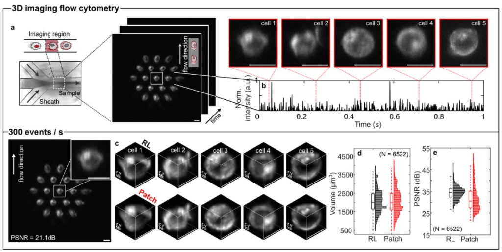
Patch deconvolution for Fourier light-field microscopy
Bin Fu, Caroline L. Jones, Daniel Heraghty, Shengbo Yang, Caitlin O’Brien-Ball, Victoria Junghans, Haowei Yang, Tuomas P.J. Knowles, Lucien E. Weiss, Ricardo A. Fernandes, Steven F. Lee
Physics-Informed Generative Model for 3D Localization Microscopy
Ofri Goldenberg, Tal Daniel, Dafei Xiao, Yael Shalev ezra, Yoav Shechtman
Frequency-dependent cellular microrheology with pyramidal atomic force microscopy probes
Erika A. Ding, Sanjay Kumar
Dual-view microscopy of single-molecule dipole orientations
Yonglei Sun, Quan Wang
Enhancing STED Microscopy via Fluorescence Lifetime Unmixing and Filtering in Two-Species SPLIT-STED
Andréanne Deschênes, Antoine Ollier, Marie Lafontaine, Albert Michaud-Gagnon, Jeffrey-Gabriel Steavan Santiague, Anthony Bilodeau, Christian Gagné, Paul De Koninck, Flavie Lavoie-Cardinal
MUFASA: A Continuous-Time Stochastic Framework for Realistic Fluorescence Microscopy Simulation
Wessim Omezzine, Sébastien Schaub, Laure Blanc-Féraud, Luca Calatroni
Self-contrastive learning enables interference-resilient and generalizable fluorescence microscopy signal detection without interference modeling
Fengdi Zhang, Ruqi Huang, Meiqian Xin, Haoran Meng, Danheng Gao, Ying Fu, Juntao Gao, Xiangyang Ji
Isotropic, aberration-corrected light sheet microscopy for rapid high-resolution imaging of cleared tissue
Mostafa Aakhte, Gesine F. Müller, Lennart Roos, Joe Li, Torben Göpel, Kurt R. Weiss, Aleyna M. Diniz, Jan Wenzel, Markus Schwaninger, Tobias Moser, Jan Huisken
Automated Registration and Clustering for Enhanced Localization Atomic Force Microscopy of Flexible Membrane Proteins
Creighton M. Lisowski, Gavin M. King, Ioan Kosztin
Label-Free Mapping of Subcellular Dynamics using Wide-field Interferometric Scattering Microscopy and Spectral Exponent Analysis
Caroline Livan Anyi, Hengze You, Huakun Li, Kheng Ling Goh, Tong Ling
Fabry-Pérot Microscopy for Improved Contrast Enhancement and 3D Cellular Imaging
Johannes Pittrich, Georg von Köller, Christoph Dillitzer, Daniel Sandner, Ellen Emken, Julia Sistermanns, Zsuzsanna Wolf, Martin Schlegel, Gregor Weirich, Reinhard Kienberger, Oliver Hayden, Hristo Iglev

Deep-learning-assisted SICM for enhanced real-time imaging of nanoscale biological dynamics
Z. Ayar, M. Penedo, B. Drake, J. Shi, S. M. Leitao, I. Krawczuk, H. Miljkovic, A. Radenovic, J. Ban, V. Cevher, G. E. Fantner
FLUID-CELL: Flow-enabled Light and Ultrastructural Imaging Device for Correlative Electron and Light Localization
Nicholas M. Rienstra, Steve Garvis, Juan C. Sanchez, Bryan Sibert, Elizabeth R. Wright
Optical Coherence Tomography with Fluorescein Optical Clearing for Transscleral Image Guidance
Robert Trout, Amit Narawane, Christian Viehland, Vahid Ownagh, Mark Draelos, Al-Hafeez Dhalla, Anthony N. Kuo, Cynthia A. Toth
A Multimodal Adaptive Optical Microscope For In Vivo Imaging from Molecules to Organisms
Tian-Ming Fu, Gaoxiang Liu, Daniel E. Milkie, Xiongtao Ruan, Frederik Görlitz, Yu Shi, Valentina Ferro, Nikita S. Divekar, Wei Wang, Harrison M. York, Velat Kilic, Matthew Mueller, Yajie Liang, Timothy A. Daugird, Maria J. Gacha-Garay, Kathryn A. Larkin, Rebecca C. Adikes, Nathaniel Harrison, Cyna Shirazinejad, Samara Williams, Jamison L. Nourse, Shu-Hsien Sheu, Liang Gao, Tongchao Li, Chandrani Mondal, Kemal Achour, Wilmene Hercule, Daniel Stabley, Kevin Emmerich, Peng Dong, David Drubin, Zhe J. Liu, David Clapham, Jeff S. Mumm, Minoru Koyama, Alison Killilea, Jose Javier Bravo-Cordero, C. Dirk Keene, Liqun Luo, Tom Kirchhausen, Medha M. Pathak, Senthil Arumugam, James K. Nunez, Ruixuan Gao, David Q. Matus, Benjamin L. Martin, Ian A. Swinburne, Eric Betzig, Wesley R. Legant, Srigokul Upadhyayula
Multiplexed Brain and Visceral Two-Photon Imaging Using a Simulation-Guided Ultrafast Three-Color Fiber Laser
Marvin Edelmann, Andreu Matamoros-Angles, Mohsin Shafiq, Mikhail Pergament, Franz X. Kärtner, Markus Glatzel
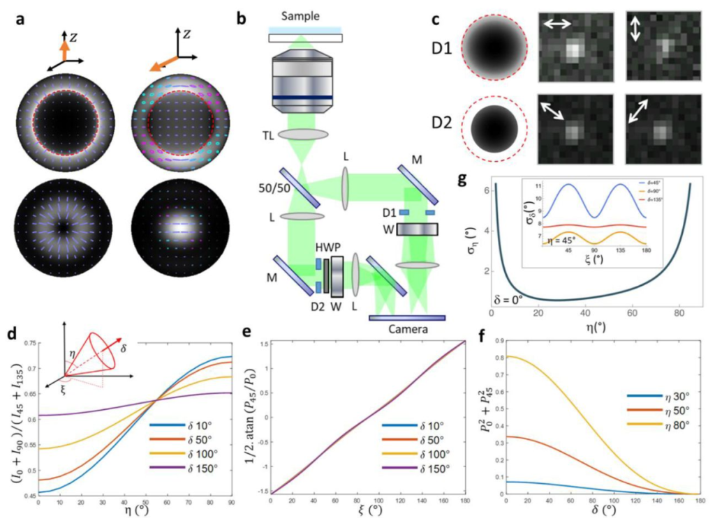
4polar3D : Single molecule 3D orientation imaging of dense actin networks using ratiometric polarization splitting
Charitra S. Senthil Kumar, Cesar A. Valades Cruz, Miguel Sison, Arturo G. Vesga, Javier Rey-Barroso, Valentina Curcio, Luis A. Alemán-Castañeda, Miguel A. Alonso, Renaud Poincloux, Manos Mavrakis, Sophie Brasselet
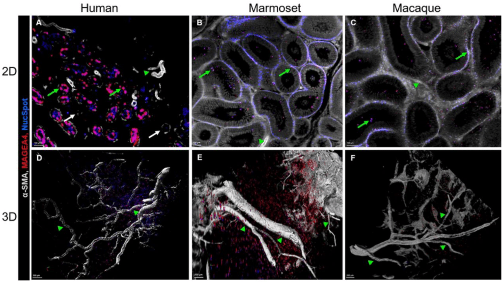
Optical tissue clearing and 3D imaging of intact primate testicular tissue: a novel technology development
Pauline Wanjiku Kibui, Sarah Weischer, Nicola von Ostau, Jochen Hess, Nils Kirschnick, Thomas Zobel, Stefan Schlatt
In situ structural analysis of mammalian cells using a 200 kV electron cryomicroscope – implications for research infrastructure
Piotr Szwedziak
Evaporation and Focus Degradation Mitigation in In-Incubator Live Cell Imaging for Capacitance Lab-on-CMOS Microsystem Calibration
Y. Gilpin, C.-Y. Lin, M. Dandin
 (No Ratings Yet)