Featured image with David Grainger
Posted by FocalPlane, on 29 August 2025
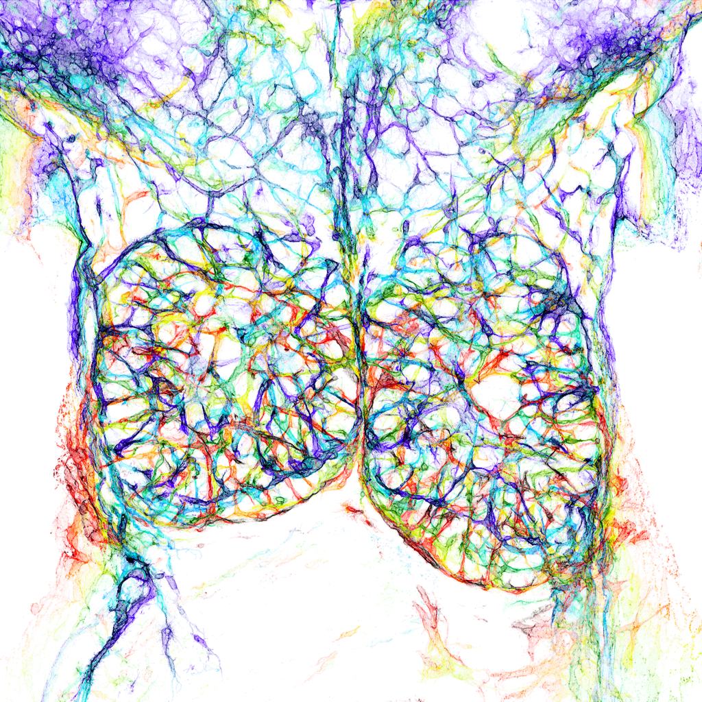
Our featured image, acquired by David Grainger, is a colour-coded depth projection of the blood endothelial cells of the E14.5 mouse embryonic thymus and surrounding structures. A 200 μm-thick vibratome section was immunostained for endomucin and imaged on a Zeiss LSM980 confocal microscope and depth-encoded using a rainbow LUT before performing a maximum intensity projection.

Discover more about David’s research
Research career so far: My research career began during my undergraduate degree in Medical Science at the University of Birmingham, where I worked with Dr Laura O’Neill and Dr Giacomo Volpe studying the epigenetic regulation of X chromosome inactivation and acute myeloid leukaemia. Their support and encouragement allowed me to move directly to a PhD with Prof. Catherine Porcher at the Weatherall Institute of Molecular Medicine (WIMM), as part of the University of Oxford, where I studied the earliest stages of blood fate acquisition to inform the development of new strategies for the directed differentiation of human pluripotent stem cells. Since finishing my PhD two years ago, I have been working with Dr Oliver Stone in the Institute of Developmental and Regenerative Medicine at the University of Oxford investigating the developmental origins of blood and lymphatic endothelial cells.
Current research: My current research as part of Ollie Stone’s research group focusses on how the developmental origin of endothelial cells impacts their considerable heterogeneity in state and function. We are studying the molecular and anatomical diversity in endothelial progenitors of the mouse embryo to determine the varied differentiation trajectories endothelial cells undertake during embryogenesis. These findings are helping us to define the origin, intermediate states and cell extrinsic signalling required to direct the differentiation of pluripotent stem cells to specialised endothelial cells. Our most recent work has identified a specialised endothelial progenitor population that directly differentiate into lymphatic endothelial cells and we are working to reproduce this in vitro. We hope this approach will allow us to identify regulators of human lymphatic endothelial cell specification, and efficiently generate them in vitro.
Favourite imaging technique/microscope: As a developmental biologist studying the mouse embryo, I am a heavy user of the laser scanning confocal microscopes. Visualising cells through immunostaining or FISH-HCR in thick tissue sections has really allowed us to map molecularly distinct cells to diverse anatomical locations and thereby better understand their fate and function within the embryo. Doing this in three dimensions with high resolution is something required for my featured image but also for many aspects of my research.
What are you most excited about in microscopy? I am excited by technologies that allow us to increasingly multiplex the number of markers we can stain and image at one time with increasing ease. The application of -omics technologies has significantly expanded the complexity of molecularly defined cell identities and states so being able to label and image this complexity in situ is very exciting.


 (No Ratings Yet)