Featured image with Denise Scuffi
Posted by FocalPlane, on 30 January 2026
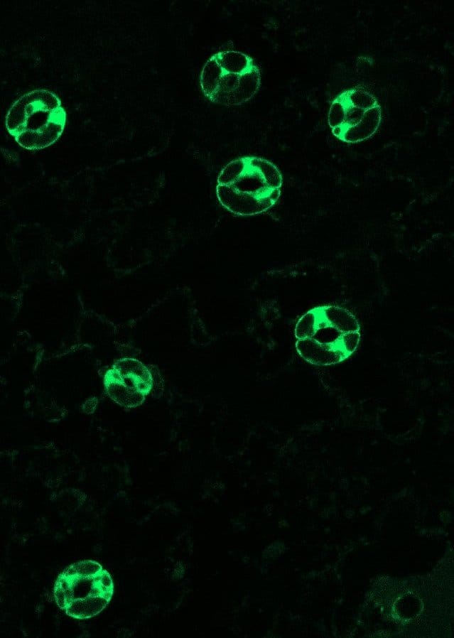
Our featured image, acquired by Denise Scuffi, shows stomata – small pores surrounded by a pair of specialised cells known as guard cells – from Arabidopsis thaliana expressing the fluorescent biosensor roGFP2-Orp1 in the cytosol, which enables in vivo monitoring of intracellular H₂O₂ dynamics. The sample consists of isolated epidermal peels obtained from the abaxial side of the leaf and floated in a buffer that promotes stomatal opening. Epidermal peels were then mounted on a confocal laser scanning microscope (CLSM). Images were acquired using a 40× objective, with the biosensor sequentially excited at 405 and 488 nm and fluorescence emission detected at 525 nm. Ratiometric images were analysed using ImageJ, and the oxidation state of the biosensor was calculated; higher oxidation levels indicate increased intracellular H₂O₂.
For additional methodological details, see: Pantaleno R, Schiel P, García-Mata C, Scuffi D. Analysis of guard cell readouts using Arabidopsis thaliana isolated epidermal peels Bio Protoc. 2024;14:e5033.
Discover more about Denise’s research.
Research career so far: I studied Biology at the National University of Mar del Plata (UNMdP) from 2004 to 2011 and completed my PhD between 2011 and 2017 at the Instituto de Investigaciones Biológicas (IIB-CONICET-UNMdP), under the supervision of Dr Carlos García-Mata and Dr Ana M. Laxalt in the ‘Signalling Mechanisms in Plants’ laboratory. I subsequently carried out postdoctoral research in the same laboratory. From the beginning of my career, my research has focused on signalling networks in plant cells. My work primarily explores the role of second messengers – such as gasotransmitters, lipids, reactive oxygen species (ROS), and calcium dynamics – in the regulation of stomatal movements in
model plant systems. In particular, I have been studying hydrogen sulfide (H₂S) signalling in stomatal movements since 2011. Our group was among the first to elucidate the H₂S signalling network promoting stomatal closure, demonstrating that H₂S actively participates in both abscisic acid- and pathogen-induced stomatal closure. To deepen these studies, it was essential to develop strong expertise in advanced microscopy and quantitative image analysis. For this purpose, I conducted two short research stays in the laboratory of Dr Markus Schwarzländer in Bonn (2016, as a PhD student) and in Münster (2021, as a visiting researcher), Germany, where I received specialized training in the use and analysis of genetically encoded fluorescent biosensors.
Current research: Currently, I am a young researcher in the ‘Signalling Mechanisms in Plants’ group led by Dr Ana M. Laxalt, where I continue to investigate plant cell signalling in collaboration with national and international research teams. In parallel, I supervise and train students pursuing academic careers and serve as a teacher at the National University of Mar del Plata (UNMdP), Argentina. My current research line focusses on the main mechanism of action of H₂S, namely persulfidation of cysteine residues in target proteins. Persulfidation is a redox-based post-translational modification that can alter protein activity, subcellular localisation, and protein-protein interactions. We are particularly interested in identifying which proteins are persulfidated and in understanding how this modification operates in guard cells under different physiological and stress conditions. To address these questions, we study the dynamics of multiple signalling molecules using chemical probes and genetically encoded biosensors based on complementary technologies.
Favourite imaging technique/microscope: My favourite technique is undoubtedly the use of fluorescent biosensors combined with CLSM. This approach allows us to monitor the in vivo dynamics of different ions and signalling molecules under physiological conditions and at distinct subcellular levels in plant cells.
What are you most excited about in microscopy?
I am deeply impressed by the power of microscopy. In biochemical sciences, the ability to directly visualise dynamic processes in vivo represents a major advance over classical biochemical approaches. Microscopy allows us to access distinct regions of the cell and observe multiple processes in real time, overcoming the limitations imposed by cellular compartmentalisation.
 (No Ratings Yet)