Imaging spotlight: Minimally invasive live tissue clearing with SeeDB-Live
Posted by Takeshi Imai, on 1 April 2026
In this paper highlight from Shigenori Inagaki and Takeshi Imai, we learn about SeeDB-Live, a methodology for applying tissue clearing to live-cell imaging.
Can you briefly describe SeeDB-Live and highlight what’s new about your methodology?
Tissue clearing is widely used for 3D fluorescence imaging of fixed (dead) samples at tissue and organ scales. The principle is very simple: the lower the index mismatch, the less light is refracted and scattered, resulting in greater transparency. However, this technique has never been applied to live cell imaging due to its toxicity. In particular, the extremely high osmolarity of the reagents has been problematic. For example, our first-generation tissue clearing agent, SeeDB, contained the saturating concentration of fructose.
In this study, we focused on high-molecular-weight chemicals instead of small molecules for index matching and found that bovine serum albumin (BSA) has extremely low osmolarity. We used BSA to adjust the refractive index of the extracellular medium to 1.363 – the average refractive index of the cytosol. The BSA-containing clearing media, SeeDB-Live, can clear live mammalian cells and tissues without affecting normal physiological functions. For example, normal neuronal activity was maintained following clearing with SeeDB-Live. Therefore, SeeDB-Live will be useful for live imaging of thick mammalian tissues ex vivo and in vivo.
What has SeeDB-Live been used for so far, and where do you see this extending?
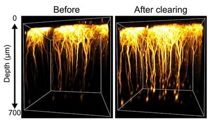
SeeDB-Live has been successfully used for a variety of live tissue samples, including cultured mammalian cells, spheroids, intestinal organoids, acute brain slices, and the mouse cerebral cortex in vivo. SeeDB-Live was useful for both structural imaging (e.g., fluorescent proteins in neurons and shadow imaging) and functional imaging (e.g., calcium and voltage imaging using genetically encoded indicators). In the brain slices, clearing enabled us to image twice as deeply for both confocal and two-photon imaging. In the mouse cerebral cortex in vivo, the fluorescence of cortical layer 5 was three times brighter. Therefore, SeeDB-Live should facilitate neurophysiology of the deeper parts of the brain.
We believe that our clearing strategy also facilitates 1-photon-based fluorescence imaging. For example, following clearing with SeeDB-Live, we successfully performed voltage imaging of neurons in thick brain slices using epifluorescence imaging at 2 kHz. We also performed epifluorescence voltage imaging of dendrites in vivo. SeeDB-Live may also be useful for super-resolution imaging, as it minimises aberrations and scattering.
Do you have any tips or tricks for researchers trying to set up and use your method?
It is important to use objective lenses designed for the refractive index of SeeDB-Live. Neither water-immersion lenses (for a refractive index of 1.33) nor silicone-immersion lenses (for a refractive index of 1.41) are perfect for SeeDB-Live (refractive index 1.363). It is strongly recommended to use a water-, silicone-, or multi-immersion objective lens with a correction collar. The correction collar should be adjusted to the refractive index of SeeDB-Live. To avoid depth-dependent spherical aberrations, the immersion medium should also have the same refractive index (1.363). We often use 2,2′-thiodiethanol or glycerol solutions for this purpose. Otherwise, even if the tissue is cleared, the image quality may not improve much.
Preparation of SeeDB-Live can be a little tricky. Different vendors’ BSA contains different levels of residual salts. Furthermore, some lots are unsuitable for SeeDB-Live, like fetal bovine serum for culture media. We have posted a step-by-step protocol using BSA from bioWORLD (fraction V, cat# 22070004). SeeDB-Live/ACSF will also soon be commercially available from Nacalai Tesque.
What are the prospects for further development?
Clearing is relatively easy for cultured cells and ex vivo samples. However, epithelial tissues are more difficult to clear, because BSA cannot penetrate the tight junction. Similarly, BSA cannot easily penetrate barrier structures in vivo. To clear the cerebral cortex in vivo, it is necessary to remove the dura mater and perfuse the cortical surface so that the BSA can diffuse into the brain.
Currently, in vivo brain clearing is based on the passive diffusion of BSA. Developing a more active delivery system for BSA in the future would further improve imaging depth.
Clearing other tissues and organs may be more challenging. We need to develop more efficient chemicals and delivery systems in future studies.
Where can people find more information?
Further information can be found in our paper and on our SeeDB Resources. The paper describes the principle of the method, the optimization strategy, and examples of its application to different types of samples. If you are interested in applying SeeDB-Live in practice, we would recommend starting with the paper and then consulting the resource page.
 (No Ratings Yet)
Get involved
Create an account or log in to post your story on FocalPlane.
More posts like this

Filter by
- NewsApply
- DiscussionsApply
- How toApply
- ToolsApply
- Case studiesApply
- InterviewsApply
- JobsApply
- EducationApply
- Blog seriesApply
- Volume EMApply
- Latin American Micro..scopistsApply
- Bio-image Analysis w..ith NapariApply
- Imaging with…Apply
- Towards Global Acces..sApply
- Latin America Bioima..gingApply
- From Zero to Qupath ..HeroApply
- Asian Microscopists ..and Cell BiologistsApply
- AIC at HHMI JaneliaApply
- Deep Learning for Bi..o-image analysisApply
- GloBIAS – updates fr..om the communityApply
- Highlights from Euro..-BioImagingApply
- LSFM seriesApply
- DIY MicroscopyApply
- View all