FocalPlane x ELMI2026 image competition: vote for your favourite
Posted by FocalPlane, on 8 June 2026
Thank you to everyone that entered our image competition with ELMI2026, we were delighted to receive so many wonderful images. Together with the ELMI2026 organising committee, we’ve come up with the following shortlist for you to choose from. You can vote using the poll at the bottom of this page.
Voting starts now and will finish on 18 June, the penultimate day of the ELMI meeting. The winner will be announced on the final day of the conference. For the latest on ELMI2026, follow the Portuguese Platform of BioImaging (PPBI) on LinkedIN, Bluesky, Instagram or X.
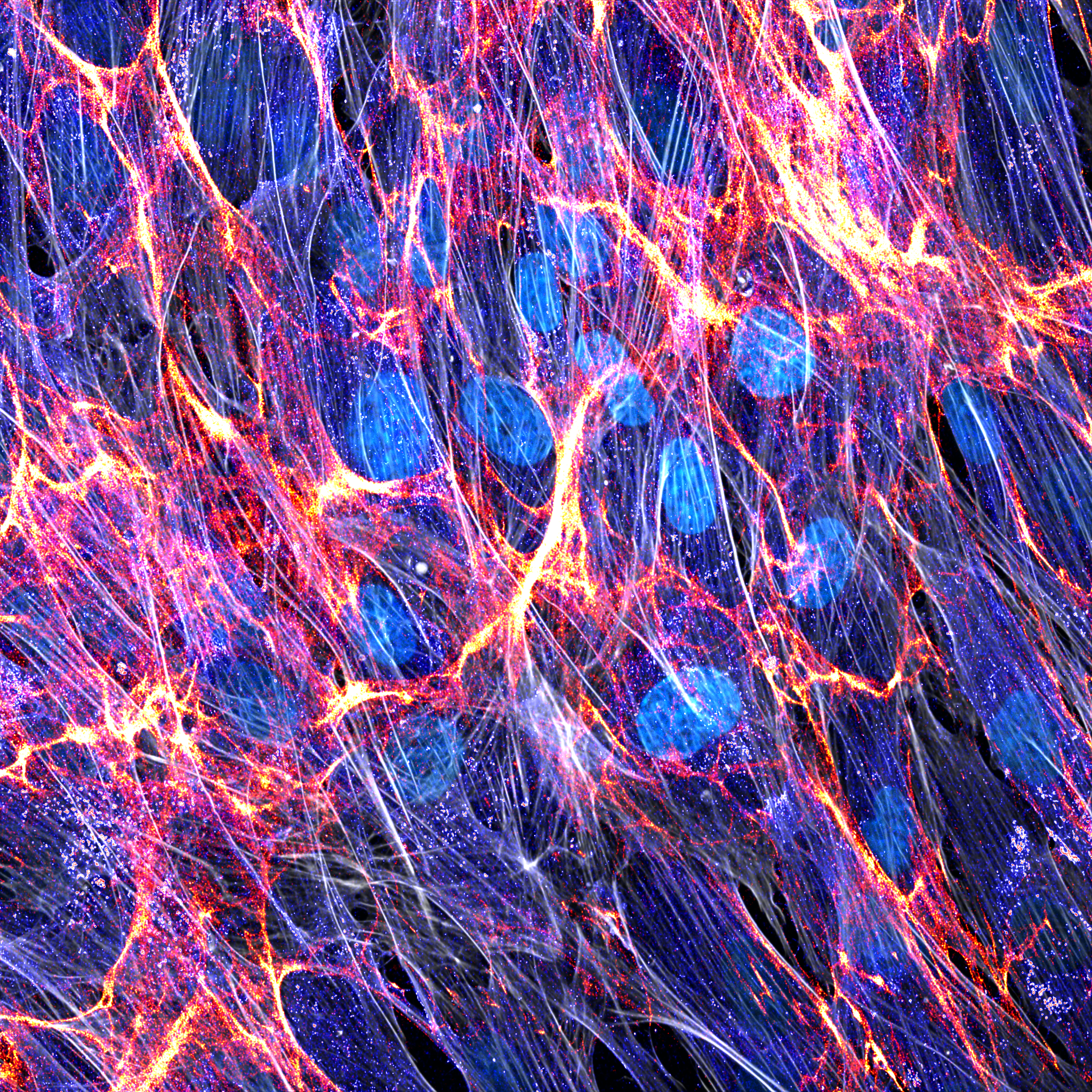
1. Marco Enriquez – Fibroblasts and ECM
The image shows telomerase-immortalized fibroblasts (TIFs) producing their own extracellular matrix. Heparan Sulfate Proteoglycan/Perlecan is shown in red and Chondroitin Sulfate Proteoglycan shown in ‘unionjack’. Phalloidin shown in gray and nuclei in cyan respectively. Imaged on an Andor Dragonfly Spinning Disk Confocal Microscope and reconstructed in Image J.
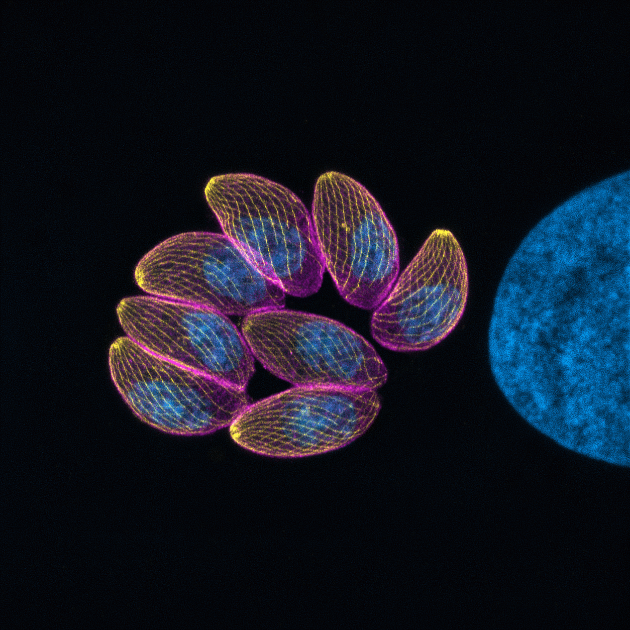
2. Ksena Longrin – Acute-stage parasites of Toxoplasma gondii
Monolayer of human foreskin fibroblasts, infected with the Type II T. gondii strain, fixed 24h post-infection, embedded in a polyacrylate gel, immunolabelled and expanded using an adapted 4-day UExM protocol. The inner membrane complex of the parasite is highlighted in magenta (anti-GAP45), microtubules in yellow (anti-beta-tubulin) and nuclei in blue (DAPI). This image is a maximum intensity projection of a z-stack of 100 optical slices, obtained using laser-scanning confocal microscope Zeiss LSM980 with a 100x objective, and then deconvoluted using Zen Blue software (Zeiss).
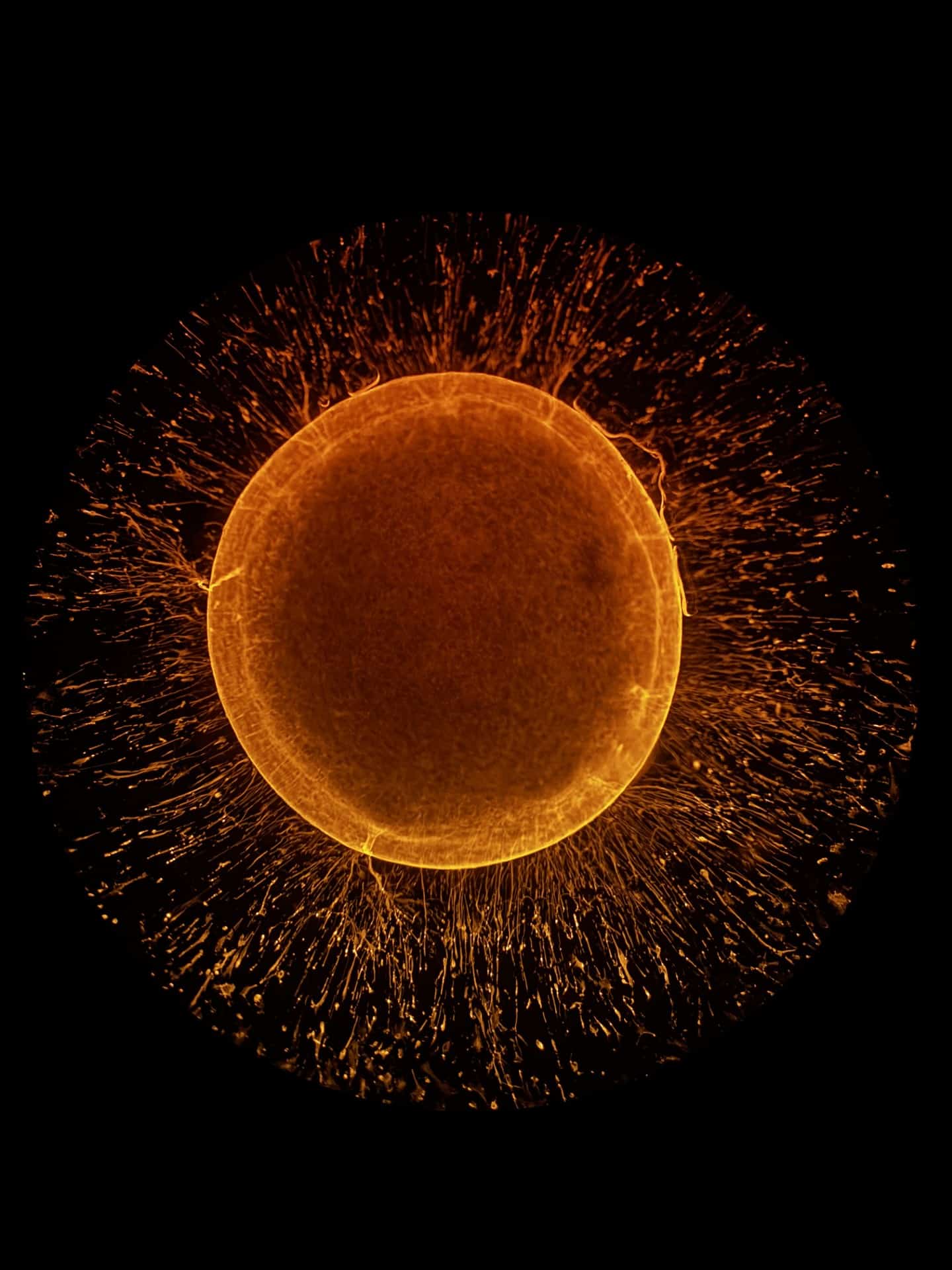
3. Mayuri Varma – Astrocytic Supernova
This image shows a human iPSC-derived cortical organoid stained for glial fibrillary acidic protein (GFAP), highlighting astrocytic structures. A dense central core is surrounded by radially organized GFAP⁺ processes that extend outward, forming a striking corona-like architecture. This spatial arrangement reflects aspects of astrocyte organization and structural scaffolding within three-dimensional neural tissue models.
The sample was imaged using laser scanning confocal microscopy.
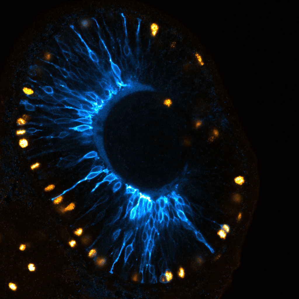
4. Maria Gorjão – Starry Eye
The image shows a 48 hpf retina from the transgenic line Tg(atoh7:GAP-GFP), which labels neurogenic progenitors. Neurons (stained for GFP) are shown in blue, while dividing cells (stained for PH3) appear in orange.
The image was acquired using an LSM 880 confocal microscope, and post-processing was performed using Fiji.
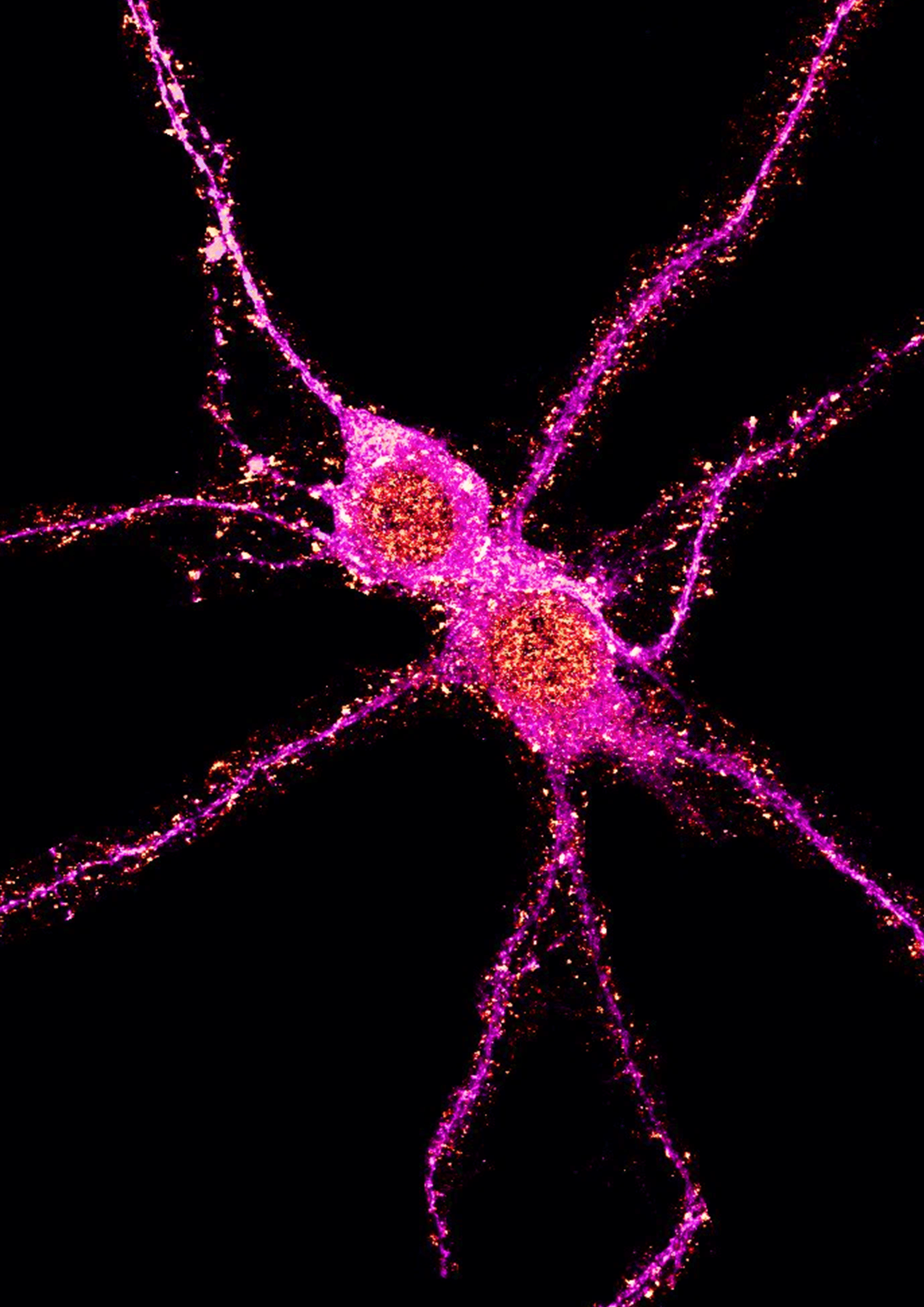
5. Jawdat Sandakly – Neuronal kiss
Mouse primary cortical neurons fixed at div14 and stained with map2, synapsin and shank2. Image was acquired with a Leica stellaris 8 DLS (University of Milan) and modified with Fiji.
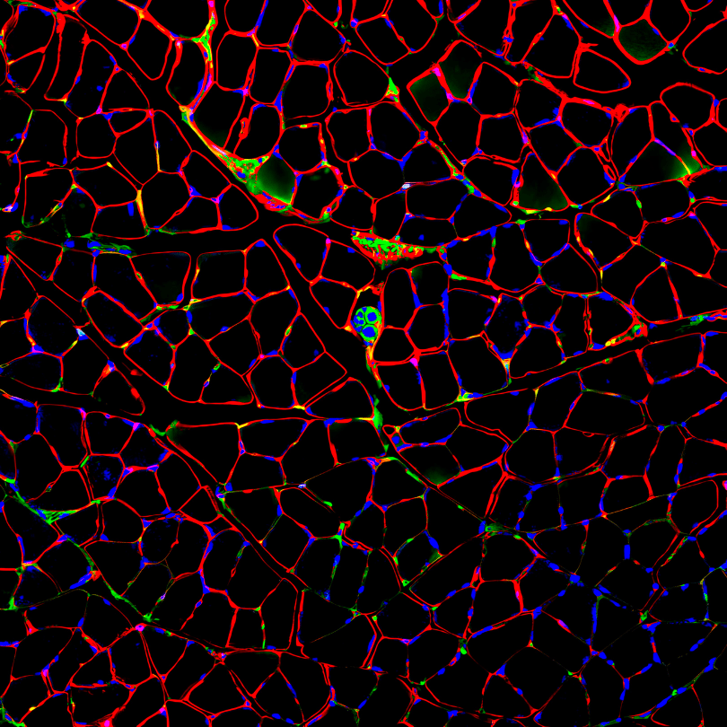
6. Rebecca Simkin – Muscle spindle
Muscle fibre basement membranes and intramuscular nerves were visualised using antibodies against laminin (red), synaptic vesicle glycoprotein 2 (SV2; green) and neurofilament M (2H3; green). Nuclei were stained with DAPI (blue). The image was acquired using a Zeiss LSM 510 confocal microscope. Colour enhancement was performed using Fiji.
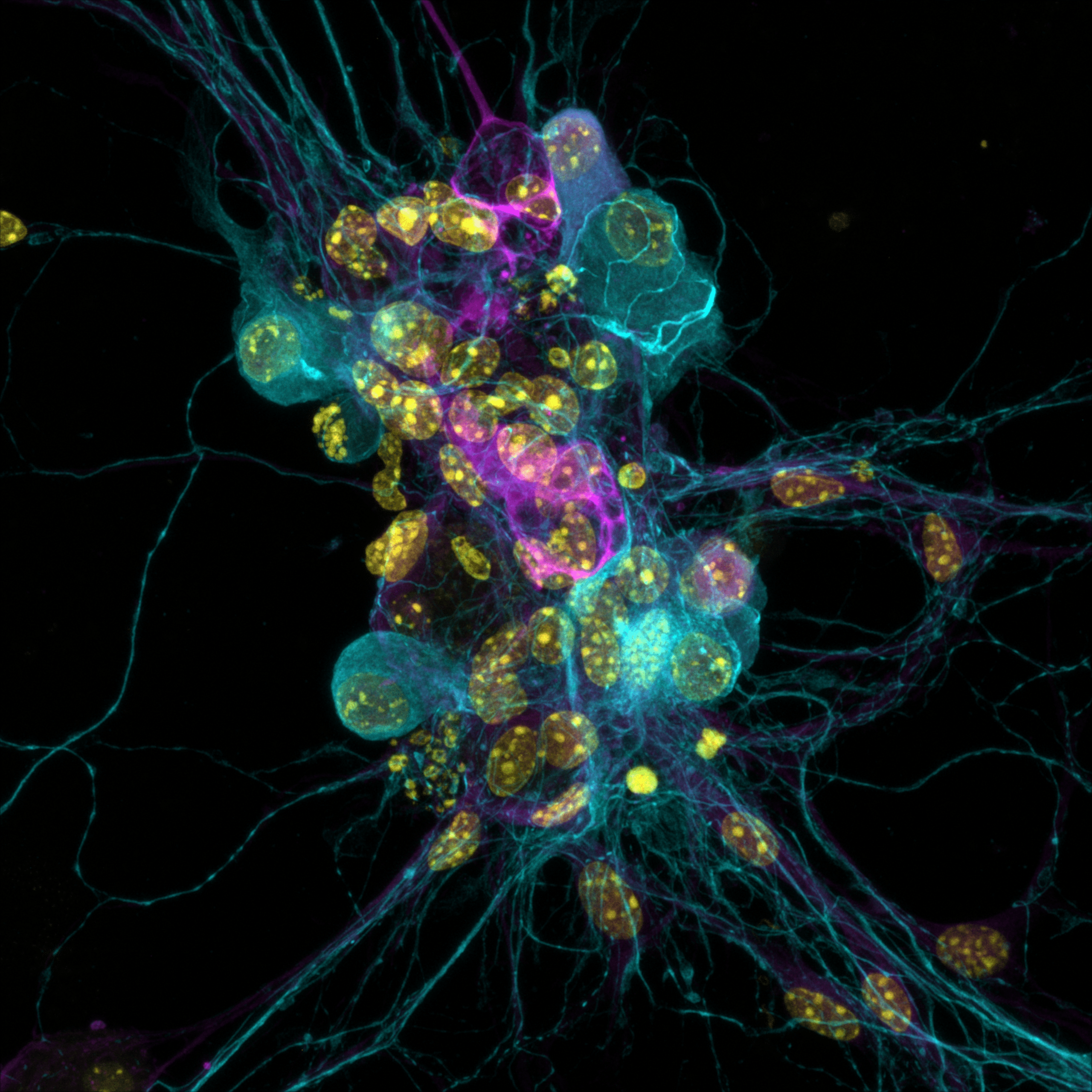
7. Pedro Campinho – Neurons and glia cells
Neuronal (Alexa-555, cyan) cell culture with glia cells (Alexa-647, magenta) and nuclear staining (DAPI, yellow). Imaging was performed on a Zeiss 980 upright confocal using airyscan-SR4Y mode and a 40x/1.2 Gly immersion objective. Zen Blue (Zeiss) was used to create a maximum intensity projection of the 99 planes (raw images).
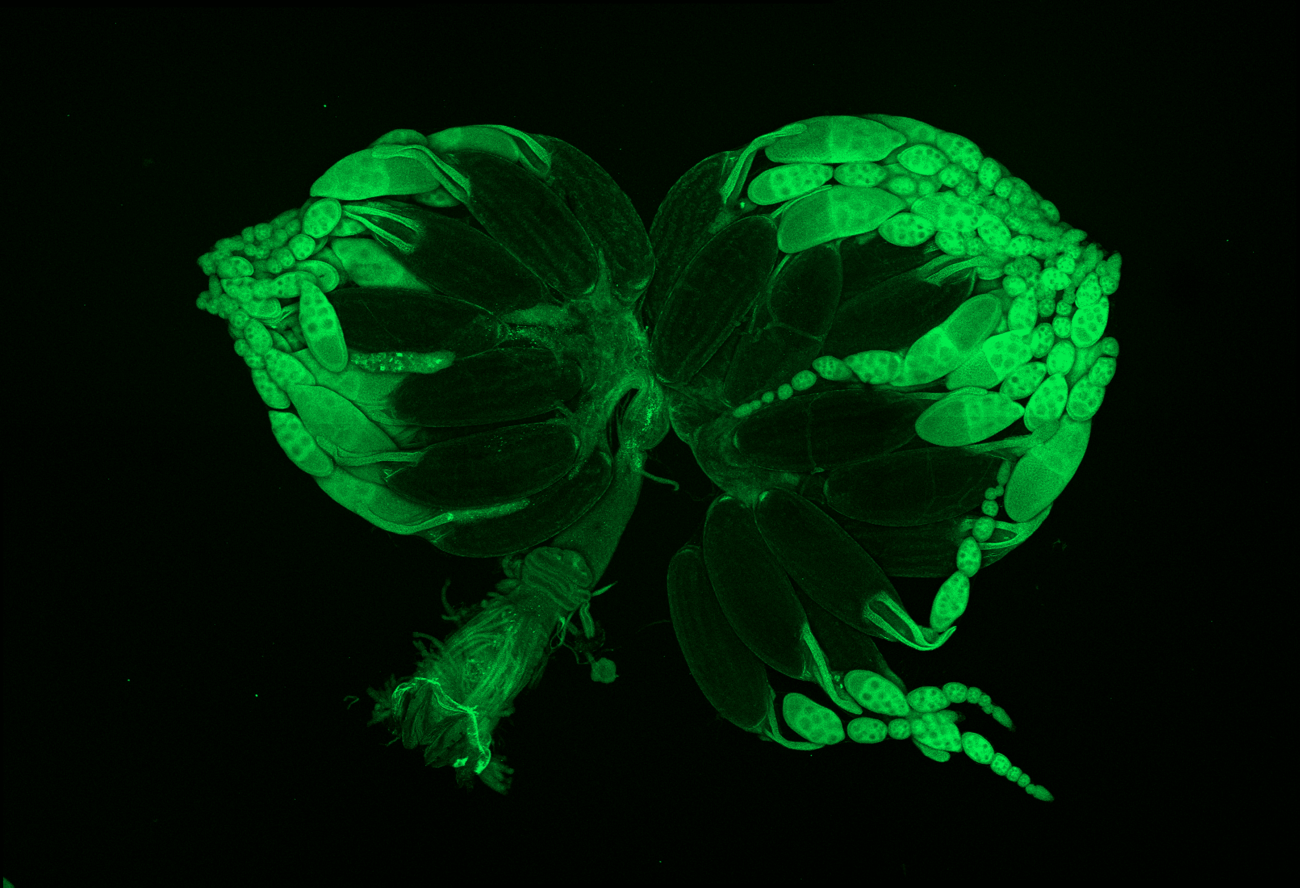
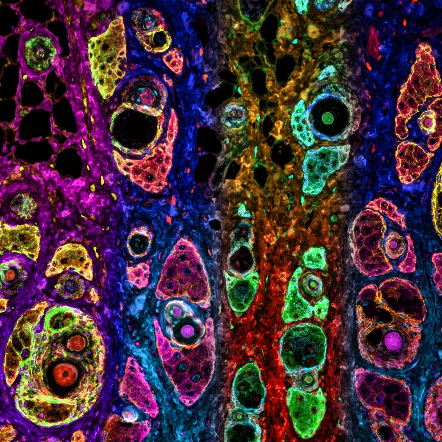
8. Saheli Roy – Flowers from a fever dream or a fly’s bursting ovaries?
The image depicts a maximum intensity projection of the reproductive system of a female Drosophila melanogaster. The ovaries are full of auto-fluorescent developing and developed eggs, because the fly had been kept in a week of ‘egg-laying deprivation’ condition. It was acquired using a Zeiss LSM 980 confocal microscope at 10X magnification, and reconstructed using Fiji.
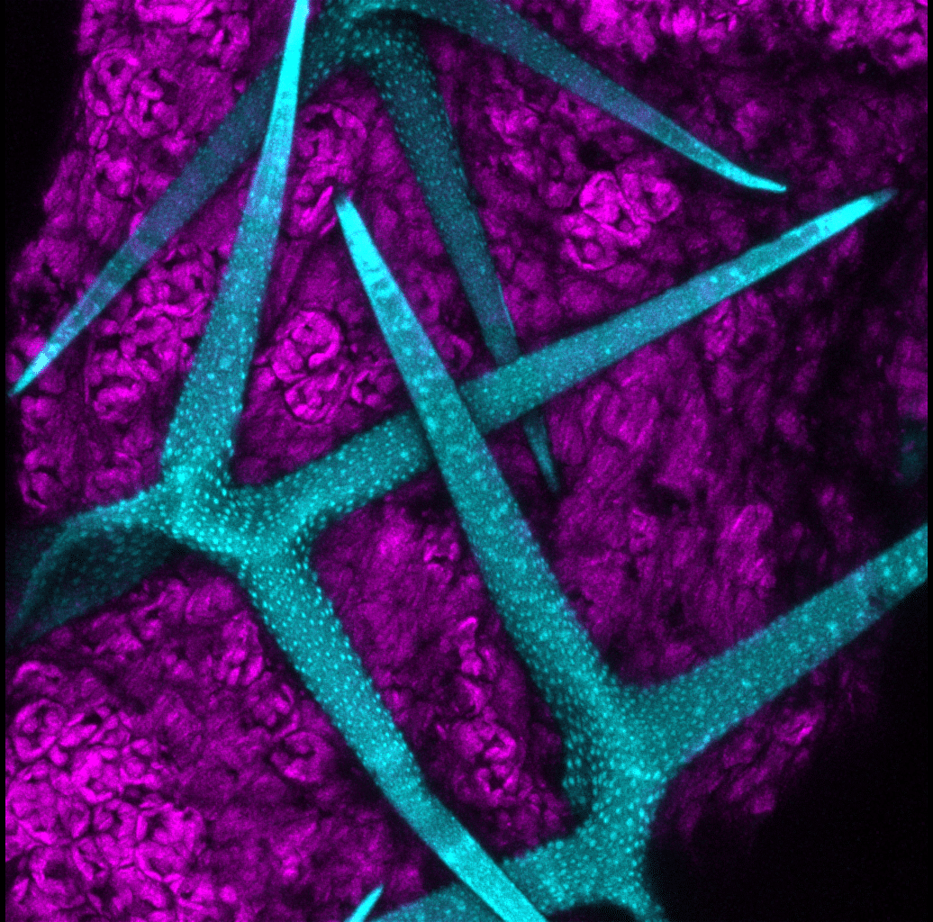
9. Luz María Fuentealba Pérez – Arabidopsis
The most widely used plant model worldwide is Arabidopsis thaliana. This organism has a small, fully sequenced genome and a short life cycle, which allows functional experiments to be conducted in a short amount of time. This image was captured just before discarding the plant that was used for an experiment; it shows autofluorescence imaged on a Leica SP8 confocal microscope using 405 nm and 633 nm lasers. Max z-projection of the image was processed in Fiji.
10. Ka Lam Nguyen – Vaginal Aurora
This image shows a multiplex fluorescence micrograph of mouse vaginal FFPE tissue stained with a 5-plex panel. The image has been processed in Adobe Photoshop to adjust hue and contrast for visual presentation.
Captured using an Olympus BX41 microscope equipped with an Olympus DP75 camera.
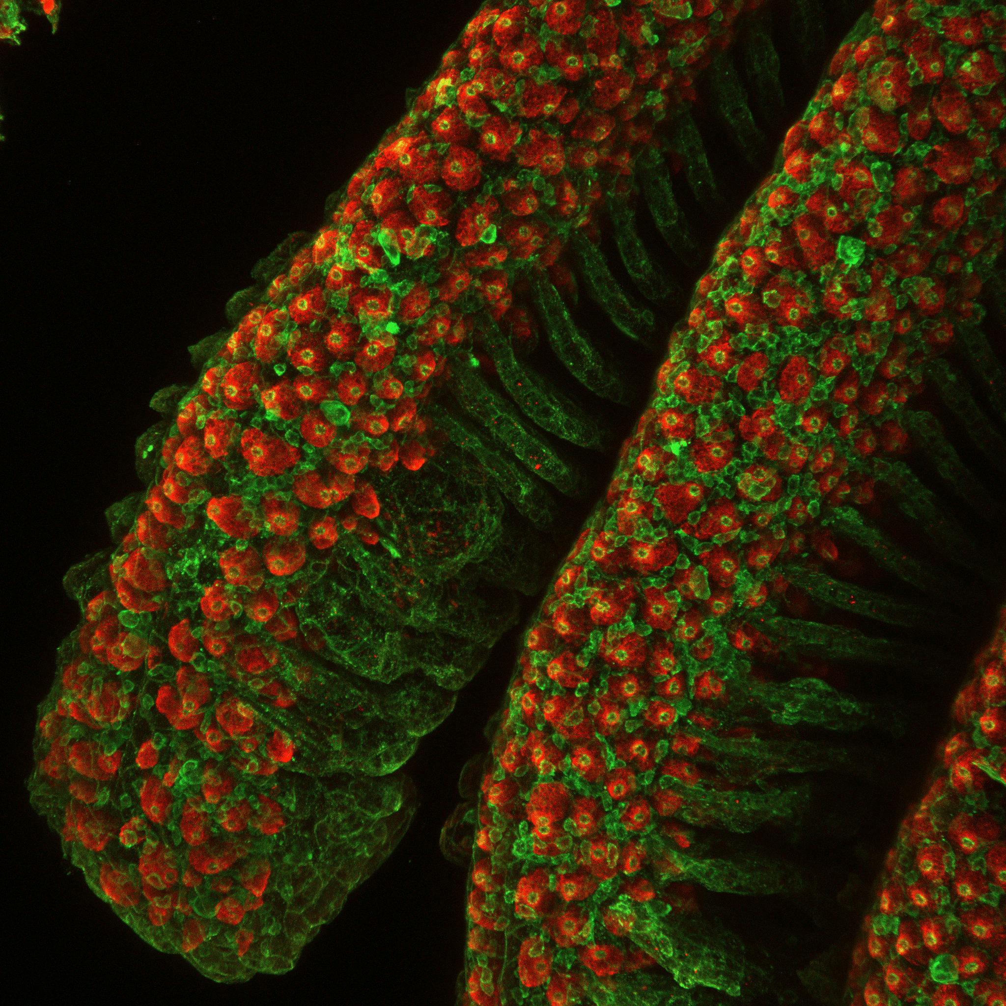
11. Shang-Wu Shih – Ionocytes in gill filaments of marine medaka
Gill tissues from marine medaka were fixed and immunostained for NKA (red) and actin (green), highlighting ionocytes in the gill filaments.
The image was acquired using a ZEISS LSM 980 confocal microscope and processed in Fiji as Z-stack projections.
Thank you for voting
 (72 votes, average: 1.00 out of 1)
Excellent
Mesmerizing image
Great work
Beautiful picture
I vote for image 3 (Astrocyte supernova). It looks superb.
Love the concept!
Love the pic
All images are equally beautiful!
Beautiful work, number 11!
Very cool ionocytes!
no.8
Beautiful
Amazing image
This image beautifully highlights the distribution of ionocytes in the gill filaments of marine medaka. The strong contrast between the red and green fluorescence provides clear visualization of cellular structures and organization. The use of confocal Z-stack projection enhances both spatial resolution and depth, making this not only a technically impressive image but also a visually striking representation of gill physiology.
No.8
Excellent work…. All the best for your immense effort… Keep it up
Vibrant
Astrocyte supernova >>
looks strikingly beautiful.
No 8
I vote for no 3
no 8
looks amazing!
I voted for No. 8 Flowers from a fever dream or a fly’s bursting ovaries. Superb
generally , all images were amazing ,,,
i vote for NO 5
Voto por el número 9
Thanks! All the entries are great.
Good Job Mayuri Varma
Voted for No 3, the energetic, classy and mesmerizing Astrocyte Supernova!
good job
Vote for mayuri Verma excellent concept keep it up
No.3
Vote for no. 11! Amazing
i found image No#5 is expressive and i vote for it
Mayank Shrivastav
I vote for number 8, mesmerising