

Microscopy preprints: bioimage analysis
Posted by FocalPlane, on 24 July 2026
Here is a curated selection of preprints published or updated recently. In this post we focus specifically on bioimage analysis and data management.
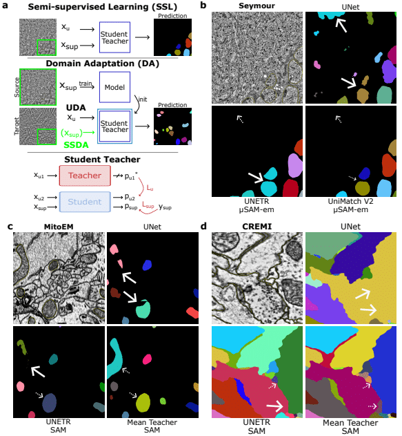
μMatch: Foundation Models for Semi-supervised Learning and Domain Adaptation in EM
Marei Freitag, Olesia Korchevaia, Luca Freckmann, Anwai Archit, Constantin Pape
SMLMFlow: Improving Structural Resolution in Single Molecule Localization Microscopy with Flow Matching
Sebastian Bauer, Luca Panconi, Inês Cunha, Emma Latron, Daniel Sage, Ruby Peters, Juliette Griffié
Apical3DTip: Elliptic Cross-section-based Reconstruction for the Embryo Initial Cell of Arabidopsis
Tomonobu Nonoyama, Zichen Kang, Yuga Hanaki, Yoshinobu Itagaki, Hikari Matsumoto, Yusuke Kimata, Satoru Tsugawa, Minako Ueda
AI-Driven Cell-Fate Prediction in Microscopy
Rita Carlota, Mario Del Rosario, Inês Cunha, Juliette Griffié, Guillaume Jacquemet, Ricardo Henriques
Higher-Order Cell Tracking Transformer
Jordão Bragantini, Ilan Theodoro, Loïc A. Royer
SparseSeg: Target-Conditioned Discovery Segmentation of Cryo-Volume Electron Microscopy Under Sparse Annotation
Bowen Shi, Yanjun Li, Qi Ouyang, Yanan Zhu
Shield-4i: A Whole-mount Multiplexed Imaging Platform for Studying Multiscale Information Flow in 3D Multicellular Systems
Ruth Hornbachner, Shayan Shamipour, Feyza Nur Arslan, Rui Fan, Max Hess, Florian Curvaia, Joel Lüthi, Andrew C. Oates, Ivan Bedzhov, Darren Gilmour, Virginie Uhlmann, Lucas Pelkmans
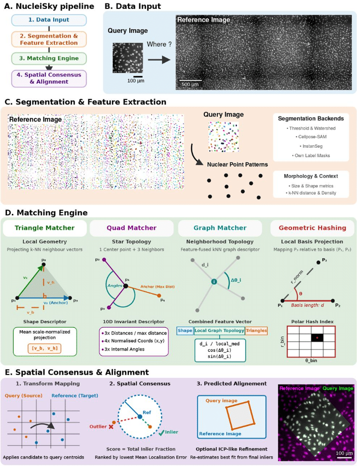
NucleiSky enables cross-scale multimodal registration of microscopy data using nuclei constellations
Iván Hidalgo Cenalmor, Adán Olguín-Olguín, Carolina Prieto, Johannes Kumra Ahnlide, Pontus Nordenfelt, Ricardo Henriques, Mario Del Rosario, Guillaume Jacquemet
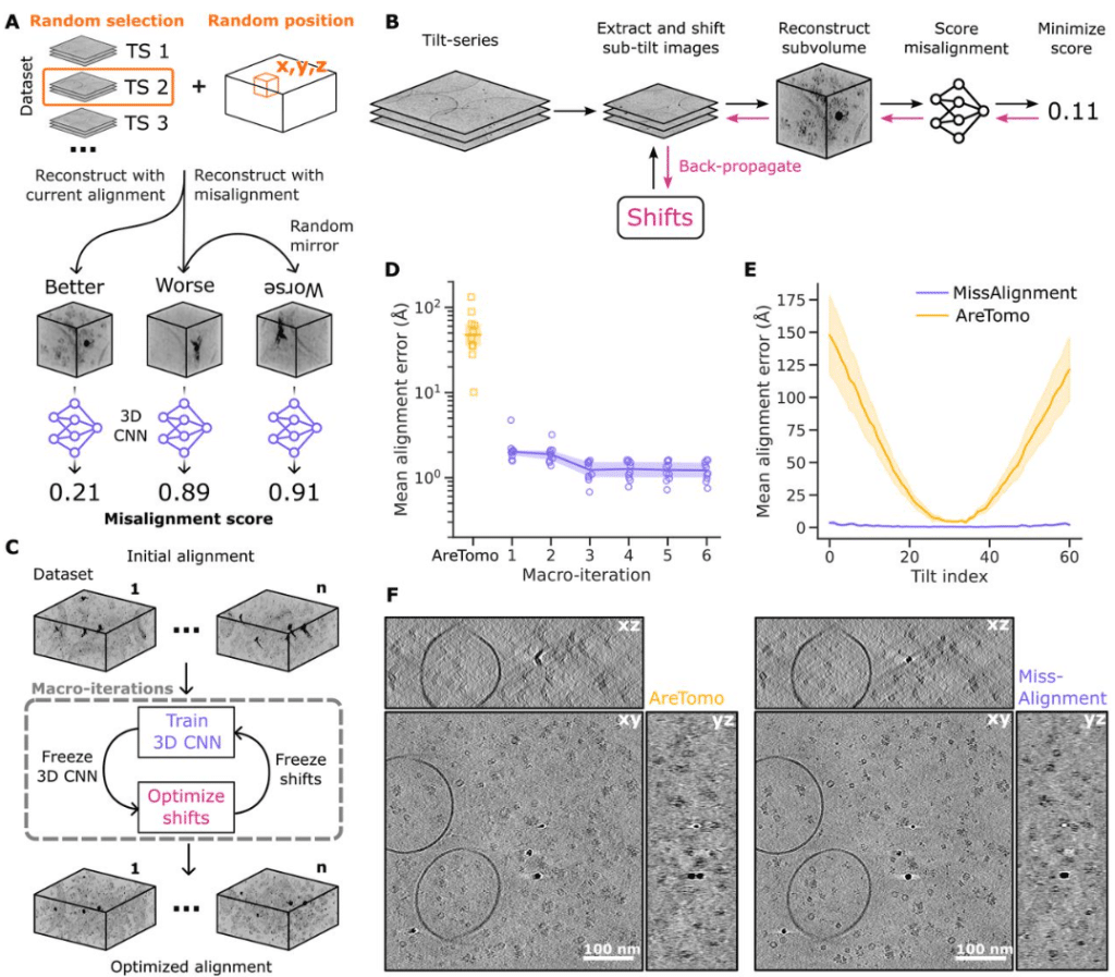
MissAlignment Teaches Itself Better Cryo-ET Tilt-Series Alignment by Making It Worse
Marten L. Chaillet, Joyce van Loenhout, Miguel R. Leung, Alister Burt, Dimitry Tegunov
iSBEM: An Open-Source Workflow for Automated ROI Targeting in Volume Electron Microscopy
Paolo Ronchi, Graham Ross, Alana Burrell, Joost de Folter, Yanneck Klenz, Nedal Darif, Fiona Young, Matthew Lawson, Jonas Albers, Tobias Pietz, Friedrich Frischknecht, Elizabeth Duke, Candice Roufosse, Lucy Collinson, Amy Strange, Yannick Schwab
CASC: Content-Aware Compaction of Sparse Microscopy Images
Gail McConnell
Vessel Spatial Analysis (VeSpA): a tool for whole slide image segmentation, morphometry, and QuPath extension
Giulia Grion, Rash Hussain, Filippo Emanuele Colella, Kirollos Roufail, Silvia Uccella, Roberta Frapolli, Cristina Matteo, Ömer Mintemur, Francesca Pennati, Salvatore Lorenzo Renne
msaGUI: Multispectral Analysis Graphical User Interface for Ratiometric Analysis and Background Correction
Grayson R. Hoy, Caitlin M. Davis
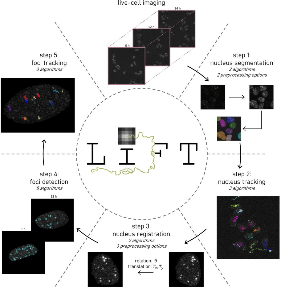
LiFT: Live foci tracking for quantitative analysis of DNA damage dynamics
Tijmen H. de Wolf, Pleun A.M. Engbers, Justine Perrin, Krijn H. van der Steen, Sam F.B. van Beuningen, Ihor Smal, Julie Nonnekens
dSTORMQuant: A Python Package for Post-Processing and Quantitative Analysis of SMLM datasets
Suraj Karki, Britta Nemeita, Anne Sophie Hammann, Sven Thoms
Suite3D: Volumetric cell detection for two-photon microscopy
Ali Haydaroğlu, Tinya Chang, Andrew Landau, Michael Krumin, Suyash Agarwal, Sam Dodgson, Liad J. Baruchin, Maria Cozan, Jingkun Guo, David Meyer, Charu Bai Reddy, Jian Zhong, Na Ji, Sylvia Schröder, Kenneth D Harris, Alipasha Vaziri, Matteo Carandini
Semi-automated reconstruction of glomerular architecture from 3D confocal microscopy data
Yoseph M Loyd, Sharon E. Chase, Mira Krendel
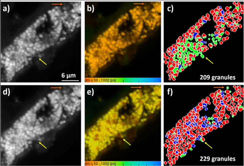
Segmentation and classification of retinal pigment granules in fluorescence lifetime imaging microscopy (FLIM) data
Maryam Ali, Hala Alhaj Ahmad, Hanan Alderzy, Martin Hammer, Rainer Heintzmann, Ondrej Stranik
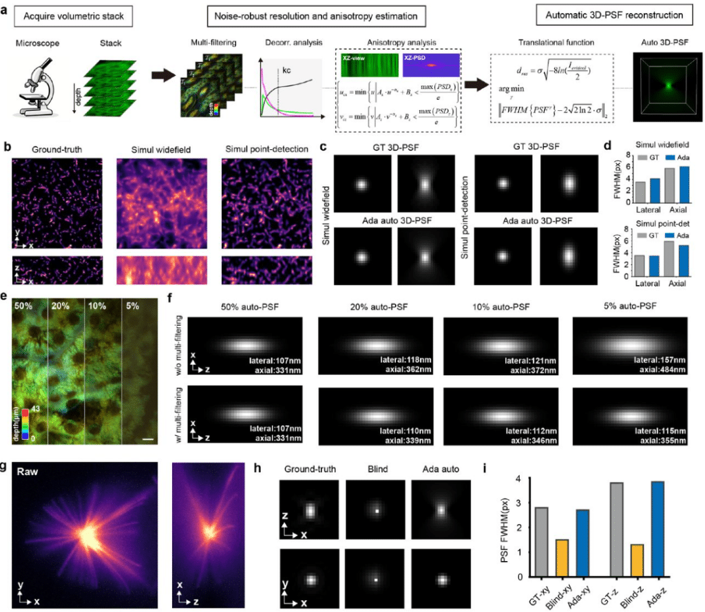
Data-adaptive three-dimensional deconvolution and evaluation for volumetric fluorescence microscopy
Yiwei Hou, Yunzhe Fu, Wenyi Wang, Ruijie Cao, Xuantao Su, Donghyun Kim, Meiqi Li, Peng Xi
MISO: A Controlled Ablation of Masking, Initialization, Sampling, and Optimization for Segmentation in Volumetric Electron Microscopy
Shanmukha Vamshi Kuruba, George Stephenson, Vignesh Kasinath
Spectral Unmixing: A modular and reproducible Python package for directed and blind spectral unmixing in multidimensional microscopy stacks
Fabrizio Musacchio, Martin Fuhrmann
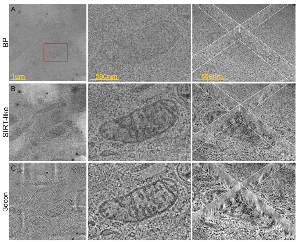
3dcon: tomogram denoising by deconvolution
Peter Kirchweger, Lev Melnikovsky, Shahar Seifer, Michael Elbaum
Penumbria: Advanced 3D cell segmentation for biomedical imaging
Laurids Stockert, Joseph Donovan, Herwig Baier
Physics-aware measurement-supervised deep learning enables point spread function inversion in soft X-ray tomography
ShaoSen Chueh, Charlotte de Ceuninck van Capelle, Leo Luo, Takashi Ishikawa, Christopher Evans, Nicola Fletcher, Mary Lopez-Perez, David Rogers, Stephen O’Connor, Conal McIntyre, Martina Donnelan, Jeremy C. Simpson, Sergey Kapishnikov
Using spIsoNet to address the preferred-orientation problem in cryoEM reconstructions
Hongcheng Fan, Yun-Tao Liu, Z. Hong Zhou