

Featured image with Saheli Roy
Posted by FocalPlane, on 3 August 2026
Our featured image, acquired by Saheli Roy, depicts a maximum intensity projection of a female Drosophila melanogaster‘s reproductive system – both ovaries, lateral and common oviducts, uterus and seminal receptacle (an organ for sperm storage that allows the female fly to control fertilization).
The ovaries are full of auto-fluorescent developing and mature eggs, because the fly had been kept for a week in ‘egg-laying deprivation’ condition, on sub-optimal substrate. Hence, we see several fully developed eggs towards the base of the ovaries, while egg chambers from earlier developmental stages are present towards the top. The image was acquired from dissecting and fixing a female fly’s reproductive system, using a Zeiss LSM 980 confocal microscope at 10X magnification, and reconstructed using FIJI.
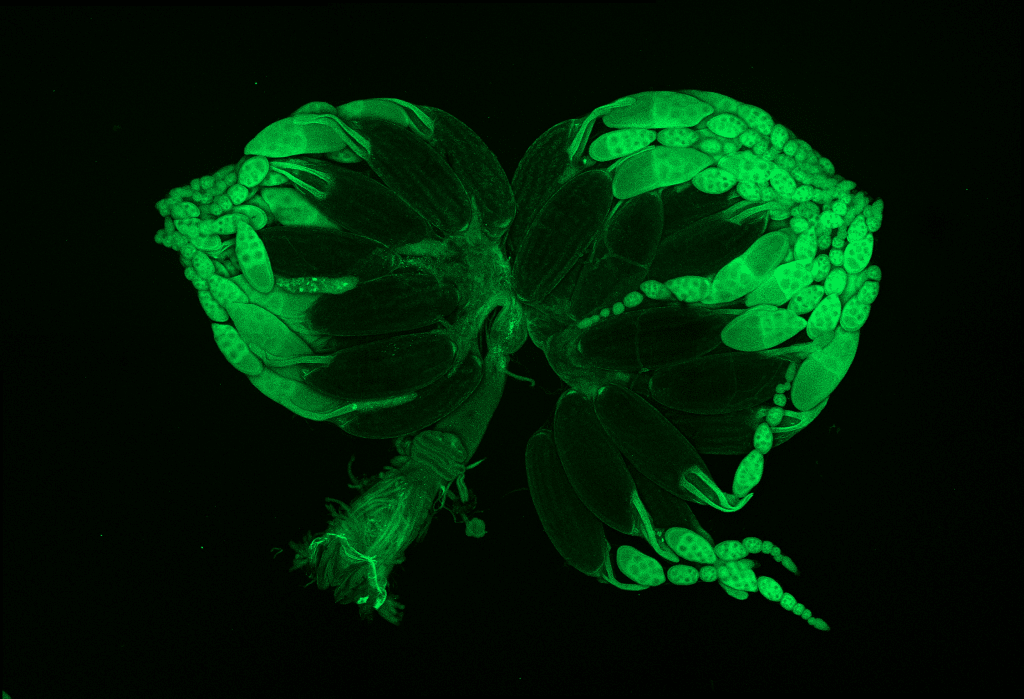
Since the fly reproductive system depicted in this image has structural similarity with other macro objects like artichokes and unbloomed lotus flowers, I titled the image as ‘Flowers from a fever dream, or a fly’s bursting ovaries?’
Read more about Saheli’s research.
Research career so far: I studied Biological Science at Presidency University, Kolkata and Jawaharlal Nehru Centre for Advanced Scientific Research (JNCASR), Bangalore, both in India. Since my undergraduate degree, I have been keen on exploring different subdisciplines of biology, hence I spent my summers pursuing research internships in labs whose focus varied from studying synthetic biology to evolution and ecology. However, being introduced to a fly lab studying chronobiology and circadian rhythms during my Master’s at JNCASR started my fascination with these tiny organisms and their rich repertoire of behaviors.
After completing my Master’s, I moved to Friedrich Miescher Institute for Biomedical Research in Switzerland to study developmental gene oscillations in another invertebrate system, Caenorhabditis elegans. However, my interest in studying behaviors only grew stronger during this experience, so in 2021 I joined the International Neuroscience Doctoral Program at Champalimaud Foundation, Lisbon, wherein I am currently pursuing my PhD in the Innate Behaviour Lab, under the supervision of Dr. Maria Luisa Vasconcelos.
Current research: My PhD project investigates hierarchy in two innate behavioural drives, using fruitflies. We are studying behavioural conflict between individual safety (i.e., defensive responses) and reproduction (i.e., species propagation). To examine this conflict, we provide looming threats while flies are engaged in egg laying, and study subsequent changes to their behaviors.
Favourite imaging technique/microscope: My favourite imaging technique is definitely calcium imaging. To me, observing neurons communicate in real-time to drive behaviour is the most satisfactory experiment one can conduct in behavioural neuroscience. But even less challenging microscopy techniques can often be quite impactful – for example, I still remember my first image acquisition of a transgenic fly brain under an epifluorescence microscope that reminded me of a starry night sky.
What are you most excited about in microscopy? I am excited about microscopy setups that allow live calcium imaging in animals while they are engaged in carrying out a behavior. I am really keen on conducting these experiments either during my PhD, or in the future!