

Microscopy preprints: bioimage analysis
Posted by FocalPlane, on 16 October 2026
Here is a curated selection of preprints published or updated recently. In this post we focus specifically on bioimage analysis and data management.
VesiScope: A stand-alone tool for the automatic detection, quantification and size analysis of GUVs from phase contrast microscopy images
Dario Cecchi, Angelo Lasala, Federica Poli, Eugenia De Remigis, Sara Moccia, Stefano Palagi
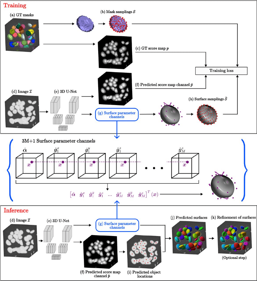
Deep Learning-Meets-Active Surfaces: A Unified Framework for Multi-Object Segmentation and Shape Analysis in 3D Fluorescence Microscopy
Quentin Rapilly, Anaïs Badoual, You-Hsuan Liu, Charles Kervrann
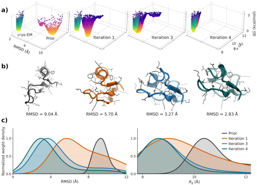
cryoWEight: Conformational ensembles from cryo-EM particle images with weighted ensemble simulations
Anupam Anand Ojha, Praveen Ranganath Prabhakar, Ioan Andricioaei, Sonya M. Hanson
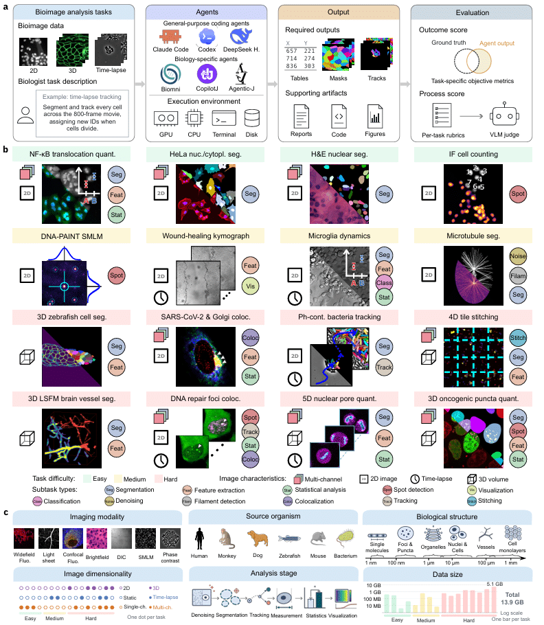
BIABench: Evaluating AI agents on real-world bioimage analysis tasks
Zixuan Pan, Davide Panzeri, Lukas Johanns, Marilin Moor, Yu Zhou, Hedi Peterson, Yiyu Shi, Jianxu Chen
Biologically grounded cell profiling across microscopy modalities
Enze Ye, Xiaoxuan Wu, Rui Peng, Wenjia Hu, Xiangyou Li, Xuefei Zhang, Mengxiao Niu, Yaorong Guo, Xinlei Sheng, Jinzhuo Wang, Liangyi Chen, He Sun
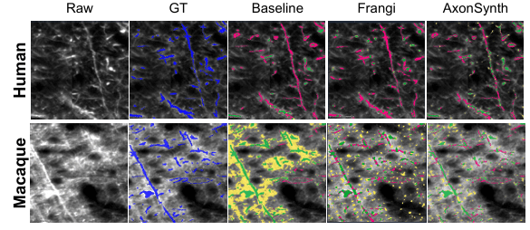
AxonSynth: Domain-Randomized Synthetic Data for Zero-Shot 3D Axon Segmentation in Light-Sheet Microscopy
Edward Gaibor, Kyriaki-Margarita Bintsi, Chiara Mauri, Carmen Luz Leiva Ureta, Zayneb Bellatif, Chiara Maffei, Wenze Li, Elizabeth Hillman, Yaël Balbastre, Anastasia Yendiki
BioAFMviewer AFM Analysis Arena A3 – an interactive software platform for automated analysis of HS-AFM imaging
Romain Amyot, Soma Yamamoto, Hiroki Konno, Clemens Franz, Holger Flechsig
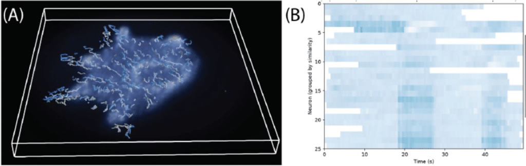
Rapid volumetric reconstruction and tracking for Fourier light-field microscopy enables real-time calcium imaging in freely behaving Hydra
Raymond Adkins, Ryan Hausen, Jamie Noss, Gerard Lemson, Jonathon Howard
Independent noise realizations enable morphologically agnostic image reconstruction in single-photon-sensitive microscopy
Lisa Cuneo, Alessandro Zunino, Laurent Le, Sofia Agostoni, Saverio Salzo, Luca Calatroni, Massimiliano Pontil, Giuseppe Vicidomini
3D-MAESTRO: A scalable, modular, portable pipeline for automated processing of large-scale volumetric brain microscopy data
Camilo Laiton, Nicholas Lusk, Yoni Browning, Mathew T. Summers, Michael Taormina, Di Wang, Joshua H. Siegle, Holly Myers, Bowen Tan, Polina Kosillo, Erica Peterson, Daphne Toglia, Anna Lakunina, Sasha Burckhardt, Mekhla Kapoor, Tim Wang, John Rohde, Galen Lynch, Shenqin Yao, Sujatha Narayan, Marcus Hooper, Sharon W. Way, Jack Waters, Bosiljka Tasic, Jayaram Chandrashekar, Adam Glaser, David Feng, Sharmishtaa Seshamani, Karel Svoboda
Pretraining and Distillation Matter More Than Architecture Family for Label-Free Single-Cell Classification
Philip Graemer, Giuseppe Di Caprio
Using a 3-step semi-automated image analysis pipeline to quantify differences in VE-cadherin junction morphology in lymphatic endothelial cells in vitro
Stephen Larson, Annabelle Feller, Catherine Whittington
OneGrow: Unified Temporal Plant Image and Mask Generation
Mike Boss, Michele Volpi, Lukas Roth
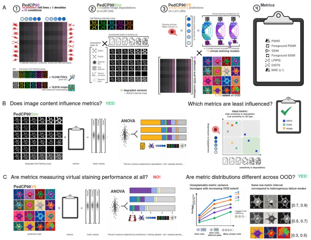
On doubting image quality assessment metrics for microscopy virtual staining
Wei-shan Li, Gregory P. Way
Quantifying performance inflation from hidden sample dependence in biomedical image classification benchmarks
Lukáš Jochymek, Markéta Vašinková, Petr Gajdš
cryoAgent: An agentic workflow for robust and adaptive end-to-end cryo-EM image processing
Daoyi Li, Shaodi Yang, Taining Pei, Chen Qi, Qian Xiao, Tongxin Niu, Yan Zhang, Yun Zhu, Fei Sun
PlanktonLake-CEREEP-A Freshwater Plankton Image Dataset with Semi-Automated Label Cleaning
Léo Déchaumet, Carine Puppo, David Carmignac, Samy Blusseau, Beatriz Marcotegui, Etienne Decencière, Gérard Lacroix, Jean-François Le Galliard
Self-Supervised Discovery of Discrete Local States in Noisy Image Sequences
Song-Chuan Zhao, Daisuke Mizuno
An Accessible Python Framework for Real-Time Magnetic Tweezers Microscope Control and Image Processing
James A. London, Abhishek K. Singh, Teague C. Svendsen, Naciye Esma Tirtom, Zachary A. Root, Richard Fishel
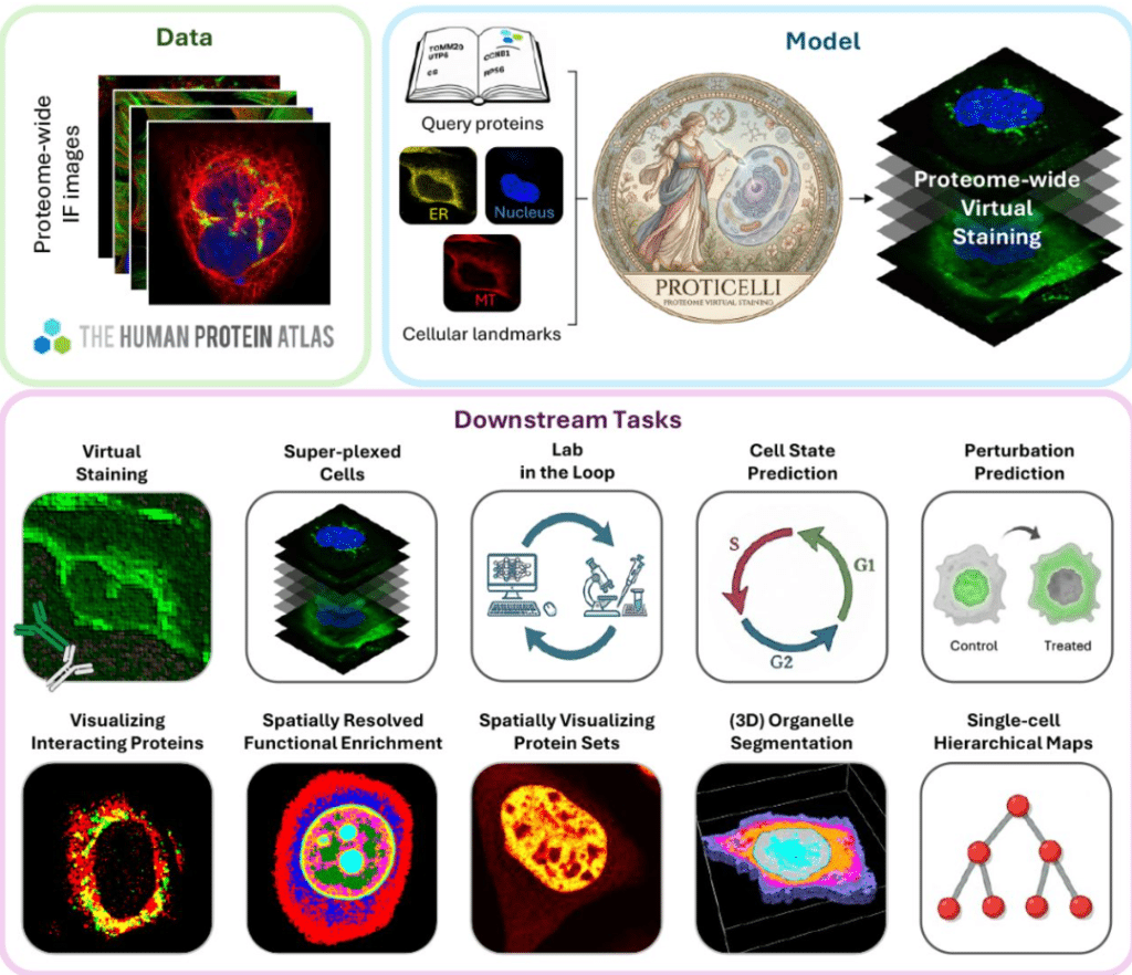
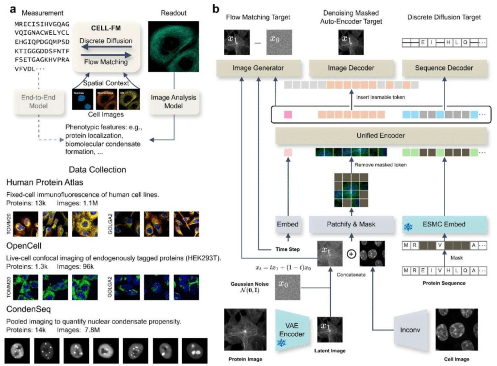
Generative machine learning unlocks the first proteome-wide image of human cells
Huangqingbo Sun, Konstantin Kahnert, Jan N. Hansen, William Leineweber, Mingyang Li, Wanyue Feng, Frederic Ballllosera Navarro, Ulrika Axelsson, Wei Ouyang, Emma Lundberg
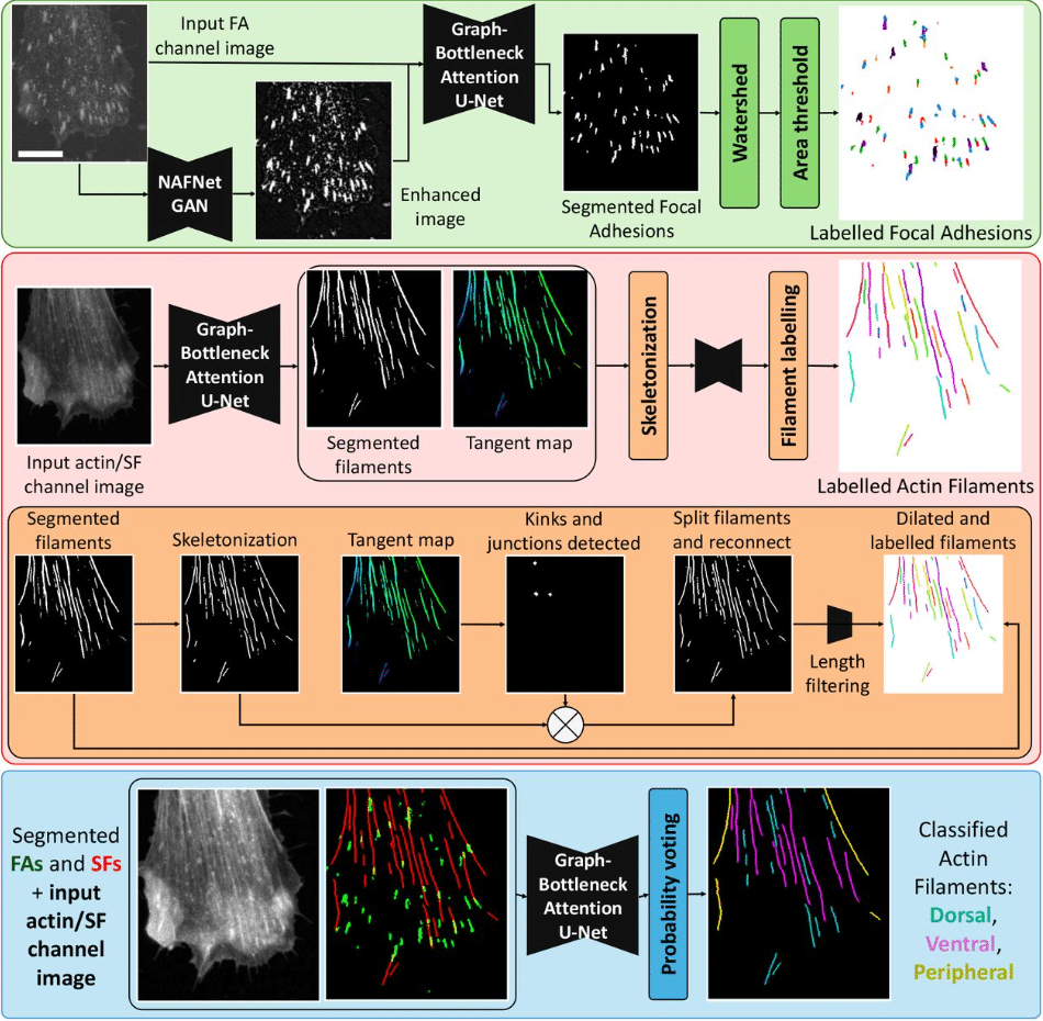
FilAdh: degradation-aware analysis of focal adhesions and actin stress fibers
Guillermo Rey-Paniagua, Carlos García-López-de-Haro, María Hernández-Martínez, Dariusz Lachowski, Arrate Muñoz-Barrutia
GENIE: a multi-agent system for closed-loop autonomous microscopy
Wilson E Nieves Vasquez, Jun Lin, Joshua Azukas, Evan Yang, Chi-Kuo Hu, Tengfei Ma, Lina Carlini
Virtual experiments bridge sequence and microscopy with generative models
Dihan Zheng, Kibeom Hong, Bo Huang
The Shape of Biological Metadata: Measuring Repository Richness with Entity-Based NLP Metrics
Maria Juliana Rodriguez-Cubillos, Tomasz Zieliński, Jason R. Swedlow, T. Ian Simpson, Andrew J. Millar
MorphCell disentangles shape and scale for 3D cellular morphology representations
Guowei Zhang, Guan Wang, Runzhou Cao, Jiansheng Guo, Shan Xu, Zeyu Yu, Yiyan Zheng, Yong He, Xuping Feng
PowerFit: An updated framework for fast rigid-body fitting into cryo-EM density maps
Anna Engel, Stefan Verhoeven, Bart Schilperoort, Rodrigo Vargas Honorato, Alexandre M.J.J. Bonvin
A general route to volumetric segmentation under annotation scarcity
Samhita Radhakrishnan, Saurabh Mathur, Aleksandr Aleksandrov, Hugo Schweke, Gerardine Silvano Gargano, Emmanuel Levy