

Featured image with Giorgia Del Cioppo
Posted by FocalPlane, on 6 November 2026
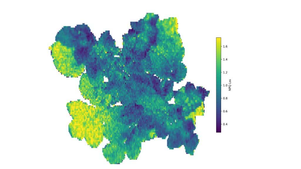
Our featured image, acquired by Giorgia Del Cioppo, shows a tomato plant (Solanum lycopersicum) subjected to drought stress, captured from a top-down perspective using pulse-amplitude-modulated (PAM) chlorophyll fluorescence imaging.
Prior to imaging, the plant underwent a standardized dark-adaptation period, followed by a quenching protocol involving actinic illumination and repeated saturating light pulses. This allows us to monitor changes in chlorophyll fluorescence and quantify non-photochemical quenching (NPQ), a sensitive digital biomarker associated with the dissipation of excess absorbed light energy as heat, which protects the photosynthetic apparatus from photodamage. In the false-colour map, NPQ values are displayed along a blue-green-yellow scale, with yellow indicating higher values. To capture the spatial heterogeneity of this response, we analysed NPQ at the pixel level using a cluster-based permutation approach, revealing localized physiological patterns across the canopy. This spatially resolved analysis showed distinct intra-canopy differences, with older leaves generally exhibiting higher NPQ values than younger tissues. Across our experiments, different stressors elicited distinct NPQ signatures. While drought-exposed plants showed elevated NPQ values compared to controls, consistent with enhanced photoprotection, salinity-exposed plants displayed a distinct peak in NPQ distribution shifted towards lower levels. This shift coincided with changes in pigment composition and likely reflects altered photosynthetic regulation.
Read more about Giorgia’s research.
Research career so far: I’ve always been fascinated by how plants perceive and respond to environmental challenges, which naturally led me toward plant physiology and phenotyping. I completed my PhD in Biology and Applied Sciences at the University of Molise (Italy), including a one-year research period at the University of Helsinki (Finland). My research has focused on plant phenotyping for stress monitoring, combining advanced imaging techniques with biochemical characterization and machine learning approaches. I am particularly interested in extracting meaningful physiological information from complex, high-dimensional datasets and translating imaging data into biologically relevant insights.
Current research: I am currently working as a Project Planner and Postdoc at the National Plant Phenotyping Infrastructure (NaPPI) at the University of Helsinki, where I contribute to the design and implementation of high-throughput phenotyping experiments across various stresses and plant species. In parallel, I am developing independent research within the CropCare project (AI-assisted potato disease monitoring – Ministry of Agriculture and Forestry), which focuses on plant-pathogen interactions. My current focus is on investigating the early stages of infection by Dickeya solani, a highly destructive bacterial pathogen of potato, with the goal of detecting physiological alterations before visible symptoms appear. Although the biological system has evolved through my work, my core scientific motivation remains the same: understanding the biological meaning of image-derived traits by linking them to the underlying physiological processes they reflect. To achieve this, I constantly integrate complementary imaging modalities to bridge localized physiological responses with whole-plant stress dynamics.
Favourite imaging technique/microscope: Chlorophyll fluorescence imaging is my favourite technique because it provides a direct, non-invasive window into photosynthetic function, which lies at the heart of plant responses to both environmental stress and disease. Unlike many other imaging approaches, it captures functional changes before structural damage becomes visible, revealing subtle physiological dynamics at the earliest stages. What I find particularly fascinating is that the more you work with chlorophyll fluorescence, the more layers of information you unlock. Every new dataset seems to reveal something unexpected, and I believe there is still enormous potential to better interpret and understand the wealth of physiological information hidden within these images.
What are you most excited about in microscopy? Looking ahead, I think one of the greatest opportunities in imaging lies in connecting information across scales, from molecular and biochemical processes to whole-plant phenotypes, through advanced imaging and data integration. By bringing these complementary layers together, we can begin to understand how processes occurring at different biological and spatial scales interact to shape the overall plant response. This will bring us closer to a predictive understanding of plant behaviour, particularly when early physiological changes are subtle and easily overlooked. At the same time, integrating imaging with machine learning is accelerating data analysis and uncovering patterns that would be nearly impossible to detect manually. However, I believe the real challenge is not simply building more accurate predictive models but ensuring that these patterns can be interpreted in a meaningful biological context. Ultimately, the greatest advances will come from using artificial intelligence to generate testable hypotheses that are grounded in plant physiology and validated experimentally.