Microscopy preprints – applications in biology
Posted by FocalPlane, on 4 October 2024
Here is a curated selection of preprints published recently. In this post, we share preprints that use microscopy tools to answer questions in biology.
Programmed cell death and stomatal density regulate anther opening in response to ambient humidity
Anna Kampová, Moritz K. Nowack, Matyáš Fendrych, Stanislav Vosolsobě
Titin-dependent biomechanical feedback tailors sarcomeres to specialised muscle functions in insects
Vincent Loreau, Wouter Koolhaas, Eunice HoYee Chan, Paul De Bossier, Nicolas Brouilly, Sabina Avosani, Aditya Sane, Christophe Pitaval, Stefanie Reiter, Nuno Miguel Luis, Pierre Mangeol, Anne C. von Philipsborn, Jean-Francois Rupprecht, Dirk Goerlich, Bianca H Habermann, Frank Schnorrer
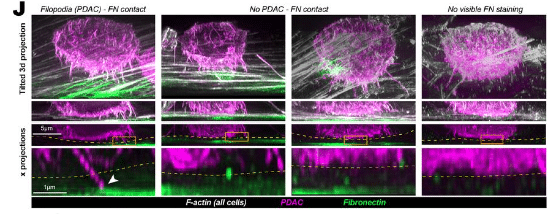
Fast label-free live imaging reveals key roles of flow dynamics and CD44-HA interaction in cancer cell arrest on endothelial monolayers
Gautier Follain, Sujan Ghimire, Joanna W. Pylvänäinen, Monika Vaitkevičiūtė, Diana Wurzinger, Camilo Guzmán, James RW Conway, Michal Dibus, Sanna Oikari, Kirsi Rilla, Marko Salmi, Johanna Ivaska, Guillaume Jacquemet
Long range mutual activation establishes Rho and Rac polarity during cell migration
Henry De Belly, Andreu Fernandez Gallen, Evelyn Strickland, Dorothy C Estrada, Patrick J Zager, Janis K Burkhardt, Herve Turlier, Orion Weiner
Mapping and engineering RNA-controlled architecture of the multiphase nucleolus
SA Quinodoz, L Jiang, AA Abu-Alfa, TJ Comi, H Zhao, Q Yu, LW Wiesner, JF Botello, A Donlic, E Soehalim, C Zorbas, L Wacheul, A Košmrlj, DLJ Lafontaine, S Klinge, CP Brangwynne
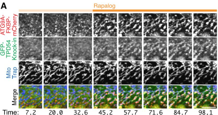
ATG9 vesicles are a subtype of intracellular nanovesicle
Mary Fesenko, Daniel J. Moore, Peyton Ewbank, Stephen J. Royle
A quantitative pipeline for whole-mount deep imaging and multiscale analysis of gastruloids
Alice Gros, Jules Vanaret, Valentin Dunsing-Eichenauer, Agathe Rostan, Philippe Roudot, Pierre-François Lenne, Léo Guignard, Sham Tlili
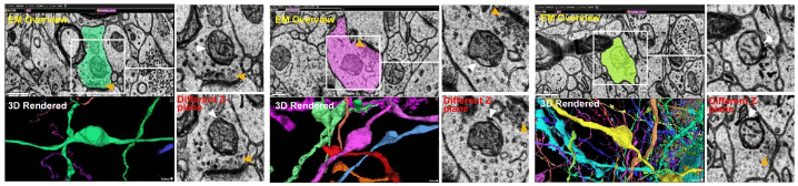
Presynapses are mitophagy pit stops that prevent axon degeneration
Wai Kit Lam, Runa S. J. Lindblom, Bridget Milky, Paris Mazzachi, Marjan Hadian-Jazi, Catharina Küng, Grace Khuu, Louise Uoselis, Thanh Ngoc Nguyen, Marvin Skulsuppaisarn, Tahnee L. Saunders, Marlene F. Schmidt, Grant Dewson, Adam I. Fogel, Cedric Bardy, Michael Lazarou
Hemifusomes and Interacting Proteolipid Nanodroplets: Formation of a Novel Cellular Organelle Complex
Amirrasoul Tavakoli, Shiqiong Hu, Seham Ebrahim, Bechara Kachar
SynPull: a novel method for studying neurodegeneration-related aggregates in synaptosomes using super-resolution microscopy
Shekhar Kedia, Emre Fertan, Yunzhao Wu, Yu P. Zhang, Georg Meisl, Jeff Y. L. Lam, Francis Wiseman, William A. McEwan, Annelies Quaegebeur, Maria Grazia Spillantini, John S. H. Danial, David Klenerman
Stress-mediated growth determines E. coli division site morphogenesis
Petr Pelech, Paula P. Navarro, Andrea Vettiger, Luke H. Chao, Christoph Allolio
Unsaturated lipids as key control points for caveola formation and disassembly
Yeping Wu, Ye-Wheen Lim, Kerrie-Ann McMahon, Nick Martel, James Rae, Harriet P. Lo, Ya Gao, Vikas Tillu, Elin Larsson, Richard Lundmark, Daniel S. Levic, Michel Bagnat, Junxian Lim, David P. Fairlie, Albert Pol, Brett M. Collins, Nicholas Ariotti, Thomas E. Hall, Robert G. Parton
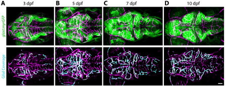
Zebrafish glial-vascular interactions progressively expand over the course of brain development
Lewis G Gall, Courtney M Stains, Moises Freitas-Andrade, Bill Z Jia, Nishi Patel, Sean G Megason, Baptiste Lacoste, Natasha Meyer O’Brown
Visualizing nuclear pore complex plasticity with Pan-Expansion Microscopy
Kimberly J. Morgan, Emma Carley, Alyssa N. Coyne, Jeffrey D. Rothstein, C. Patrick Lusk, Megan C. King
Three-color single-molecule localization microscopy in chromatin
Nicolas Acosta, Ruyi Gong, Yuanzhe Su, Jane Frederick, Karla Medina, Wing Shun Li, Kiana Mohammadian, Luay Almassalha, Geng Wang, Vadim Backman
Expansion microscopy of axonemal dyneins in islet primary cilia
Xinhang Dong, Jung Hoon Cho, Jing Hughes
Optimized expansion microscopy reveals species-specific spindle microtubule organization in Xenopus egg extracts
Gabriel Guilloux, Maiko Kitaoka, Karel Mocaer, Claire Heichette, Laurence Duchesne, Rebecca Heald, Thierry Pecot, Romain Gibeaux
Under or Over? Tracing Complex DNA Structures with High Resolution Atomic Force Microscopy
Elizabeth P. Holmes, Max C. Gamill, James I. Provan, Laura Wiggins, Renáta Rusková, Sylvia Whittle, Thomas E. Catley, Kavit H. S. Main, Neil Shephard, Helen. E. Bryant, Neville S. Gilhooly, Agnieszka Gambus, Dušan Račko, Sean D. Colloms, Alice L. B. Pyne
Detailed Colocalization Analysis of A- and B-type Nuclear Lamins: a Workflow Using Super-Resolution STED Microscopy and Deconvolution
Merel Stiekema, Owen N. Gibson, Rogier J.A. Veltrop, Frans C.S. Ramaekers, Jos L.V. Broers, Marc A.M.J. van Zandvoort
High-content microscopy and machine learning characterize a cell morphology signature of NF1 genotype in Schwann cells
Jenna Tomkinson, Cameron Mattson, Michelle Mattson-Hoss, Herb Sarnoff, Stephanie J. Bouley, James A. Walker, Gregory P. Way
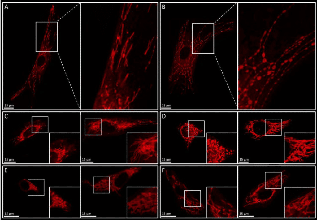
The curse of the red pearl: a fibroblast specific pearl-necklace mitochondrial phenotype caused by phototoxicity
Irene MGM Hemel, Kèvin Knoops, Carmen López-Iglesias, Mike Gerards
Examination of Lipid Distributions in Hydrogel-Expanded Mouse Brain Tissue Using Imaging Mass Spectrometry
Jacob M. Samuel, Tingting Yan, Zhongling Liang, Boone M. Prentice
Traction force generation in motile malaria parasites is modulated by the Plasmodium adhesin TLP
Johanna Ripp, Dimitri Probst, Mirko Singer, Ulrich Sebastian Schwarz, Friedrich Frischknecht
PIP2 promotes the incorporation of CD43, PSGL-1 and CD44 into nascent HIV-1 particles
Ricardo de Souza Cardoso, Tomoyuki Murakami, Binyamin Jacobovitz, Sarah L. Veatch, Akira Ono
Application of cryo-FIB-SEM for investigating organelle ultrastructure in guard cells of higher plants
Bastian Leander Franzisky, Xudong Zhang, Claus Jakob Burkhardt, Endre Majorovits, Eric Hummel, Andreas Schertel, Christoph-Martin Geilfus, Christian Zörb
Highly dynamic mechanical transitions in embryonic cell populations during Drosophila gastrulation
Juan Manuel Gomez, Carlo Bevilacqua, Abhisha Thayambath, Maria Leptin, Julio M Belmonte, Robert Prevedel
Ultrasound-activated microbubbles mediate F-actin disruptions and endothelial gap formation during sonoporation
Bram Meijlink, H. Rhodé van der Kooij, Yuchen Wang, Hongchen Li, Stephan Huveneers, Klazina Kooiman
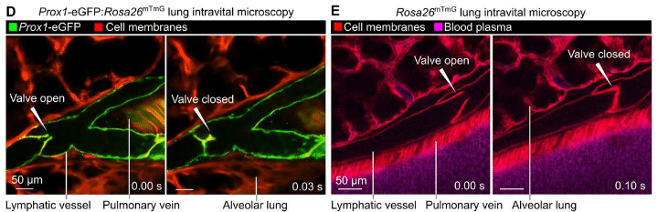
Intravital imaging of pulmonary lymphatics in inflammation and metastatic cancer
Simon J. Cleary, Longhui Qiu, Yurim Seo, Peter Baluk, Dan Liu, Nina K. Serwas, Jason G. Cyster, Donald M. McDonald, Matthew F. Krummel, Mark R. Looney
Expansion microscopy allows quantitative characterisation of structural organisation of platelet aggregates
Emma L. Faulkner, Jeremy A. Pike, Evelyn Garlick, Robert K. Neely, Iain B. Styles, Stephen P. Watson, Natalie S. Poulter, Steven G. Thomas
Two-dimensional condensates of HRS drive the assembly of flat clathrin lattices on endosomes
Markku Hakala, Satish Babu Moparthi, Iva Ganeva, Cesar Bernat-Silvestre, Javier Espadas, Wanda Kukulski, Stephane Vassilopoulos, Marko Kaksonen, Aurelien Roux
Extracellular filaments revealed by affinity capture cryo-electron tomography
Leeya Engel, Magda Zaoralová, Momei Zhou, Alexander R. Dunn, Stefan L. Oliver
In situ quantification of ribosome number by electron tomography
Mounir El Hankouri, Marco Nousch, Thomas Müller-Reichert, Gunar Fabig
NuMA is a mitotic adaptor protein that activates dynein and connects it to microtubule minus ends
Sabina Colombo, Christel Michel, Silvia Speroni, Felix Ruhnow, Maria Gili, Claudia Brito, Thomas Surrey
Live-cell imaging and CLEM reveal the existence of ACTN4-dependent ruffle-edge lamellipodia acting as a novel mode of cell migration
Haruka Morishita, Katsuhisa Kawai, Youhei Egami, Kazufumi Honda, Nobukazu Araki
3D nanoscale architecture of the respiratory epithelium reveals motile cilia-rootlets-mitochondria axis of communication
Aaran Vijayakumaran, Christopher Godbehere, Analle Abuammar, Sophia Y. Breusegem, Leah R. Hurst, Nobuhiro Morone, Jaime Llodra, Melis T. Dalbay, Niaj M. Tanvir, K. MacLellan-Gibson, Chris O’Callaghan, Esben Lorentzen, CellMap Project Team, FIB-SEM Technology, Andrew J. Murray, Kedar Narayan, Vito Mennella
 (No Ratings Yet)
Get involved
Create an account or log in to post your story on FocalPlane.
More posts like this

Filter by
- NewsApply
- DiscussionsApply
- How toApply
- ToolsApply
- Case studiesApply
- InterviewsApply
- JobsApply
- EducationApply
- Blog seriesApply
- WAMBIAN: West Africa.. in FocusApply
- Volume EMApply
- Latin American Micro..scopistsApply
- Bio-image Analysis w..ith NapariApply
- Imaging with…Apply
- Towards Global Acces..sApply
- Latin America Bioima..gingApply
- From Zero to Qupath ..HeroApply
- Asian Microscopists ..and Cell BiologistsApply
- AIC at HHMI JaneliaApply
- Deep Learning for Bi..o-image analysisApply
- GloBIAS – updates fr..om the communityApply
- Highlights from Euro..-BioImagingApply
- LSFM seriesApply
- DIY MicroscopyApply
- View all