Featured image with Md Hashim Reza
Posted by FocalPlane, on 22 November 2024
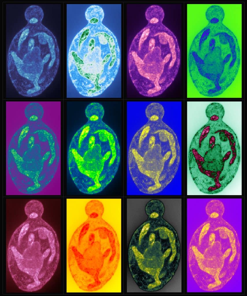
Our featured image, prepared by Md Hashim Reza, is an Andy Warhol-inspired illustration of a Candida albicans cell, post-expansion and labelled with BodipyTR, highlighting features of internal membrane organisation. The image is a maximum-intensity projection and was captured using a Nikon CSU-W1 SORA spinning disk confocal microscope.

Find out more about Md Hashim Reza’s research below:
Research career so far: My first research experience was during the final year of my undergraduate studies in the laboratory of Prof. Yogesh Singh at the Institute of Genomics and Integrative Biology (IGIB), New Delhi, India where I worked on the purification of protein kinases from Mycobacterium. During my Masters, I worked on understanding the diversity of polyketide synthases from lichens, in the laboratory of the late Prof. Bharat B Chattoo at Genome Research Centre (GRC), Vadodara, India. I continued working in his lab together with the late Prof. Johannes Manjrekar on a plant pathogen, Magnaporthe oryzae for my PhD. Combining genetics, biochemistry, and microscopy, I studied the role of magnesium transporters in the pathogen and showed how these play a role in the pathogenic life cycle of the rice blast fungus. Then I moved to Prof. Kaustuv Sanyal’s laboratory at Jawaharlal Nehru Centre for Advanced Scientific Research (JNCASR), Bengaluru, India and I am working as a postdoctoral fellow on various aspects of cell cycle progression in human fungal pathogens.
Current research: At present, I am investigating the non-canonical function of an autophagy protein, Atg11 in mediating high-fidelity chromosome segregation in the budding yeast, Saccharomyces cerevisiae. Our complementary approaches of yeast genetics, biochemistry, confocal microscopy, and single-molecule tracking assay suggest a role of Atg11 at the spindle pole bodies (SPBs) in facilitating astral microtubule dynamics crucial for proper spindle positioning and alignment.
Favourite imaging technique/microscope: Right from my PhD, I have used a confocal system from Zeiss. I have been trained on LSM700, 880, and 980. I enjoyed working on the LSM980 Airyscan confocal microscope during my stay in Dr Gautam Dey’s lab at EMBL, Heidelberg, Germany.
What are you most excited about in microscopy? The first time I saw the dynamics of a vacuolar transporter with GFP tagging in Magnaporthe oryzae, I was amazed. Over the years, a rapid evolution of imaging techniques has facilitated an improvement in resolution. The idea of enhancing the resolution by expansion microscopy, making it available to most laboratories, is remarkable and is gaining lots of attraction to answer key questions in the field. Recently, my work involved collaborating with Dr. Gunjan Mehta’s lab from the Indian Institute of Technology, Hyderabad where we could show for the first time that Atg11, an autophagy protein, shows dynamic localization at the SPBs using single-molecule tracking assay. I’m excited by these advancements that help address and validate novel observations.
Hashim’s image was acquired as part of his research investigating the ultrastructure of pathogenic budding yeast species by expansion microscopy, which was published in Journal of Cell Science’s Special Issue: Imaging Cell Architecture and Dynamics . Hashim shared this research in our FocalPlane features… webinar earlier in this month. You can view the recording below:


 (No Ratings Yet)