Microscopy preprints – applications in biology
Posted by FocalPlane, on 10 January 2025
Here is a curated selection of preprints published recently. In this post, we share preprints that use microscopy tools to answer questions in biology.
Smc5/6 association with microtubules controls dynamic pericentromeric chromatin folding
Ànnia Carré-Simon, Renaud Batrin, Sarah Isler, Henrik Dahl Pinholt, Timothy Földes, Guillaume Laflamme, Maria Barbi, Leonid Mirny, Damien D’Amours, Emmanuelle Fabre
Fishnet mesh of centrin-Sfi1 drives ultrafast calcium-activated contraction of the giant cell Spirostomum ambiguum
Joseph Lannan, Carlos Floyd, L. X. Xu, Connie Yan, Wallace F. Marshall, Surirayanarayanan Vaikuntanathan, Aaron R. Dinner, Jerry E. Honts, Saad Bhamla, Mary Williard Elting
Regulation of bicoid mRNA throughout oogenesis and early embryogenesis impacts protein gradient formation
T. Athilingam, E.L. Wilby, P. Bensidoun, A. Trullo, M. Verbrugghe, M. Lagha, T.E. Saunders, T.T. Weil
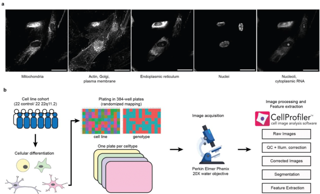
Combining NeuroPainting with transcriptomics reveals cell-type-specific morphological and molecular signatures of the 22q11.2 deletion
Matthew Tegtmeyer, Dhara Liyanage, Yu Han, Kathryn B. Hebert, Ruifan Pei, Gregory P. Way, Pearl V. Ryder, Derek Hawes, Callum Tromans-Coia, Beth A. Cimini, Anne E. Carpenter, Shantanu Singh, Ralda Nehme
Ice gliding diatoms establish record-low temperature limit for motility in a eukaryotic cell
Qing Zhang, Hope T. Leng, Hongquan Li, Kevin R. Arrigo, Manu Prakash
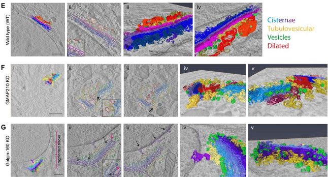
Golgins support extracellular matrix secretion by collectively maintaining the Golgi structure-function relationship
George Thompson, Anna Hoyle, Philip A. Lewis, M. Esther Prada-Sanchez, Joe Swift, Kate Heesom, Martin Lowe, David Stephens, Nicola Stevenson
Strikingly different neurotransmitter release strategies in dopaminergic subclasses
Ana Dorrego-Rivas, Darren J. Byrne, Yunyi Liu, Menghon Cheah, Ceren Arslan, Marcela Lipovsek, Marc C. Ford, Matthew S. Grubb

Automated segmentation of soft X-ray tomography: native cellular structure with sub-micron resolution at high throughput for whole-cell quantitative imaging in yeast
Jianhua Chen, Mary Mirvis, Axel Ekman, Bieke Vanslembrouck, Mark Le Gros, Carolyn Larabell, Wallace F. Marshall
Quiescent neural stem cells transiently become ‘neurons’ to coordinate reactivation
Laura-Yvonne Gherghina, Jocelyn L.Y. Tang, Andrea H. Brand
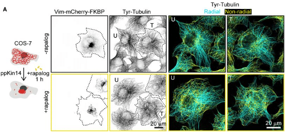
Optogenetic and chemical genetic tools for rapid repositioning of vimentin intermediate filaments
Milena Pasolli, Joyce C. M. Meiring, James P. Conboy, Gijsje H. Koenderink, Anna Akhmanova
Revealing the Ultrastructure of Live Candida albicans using Stimulated Emission Depletion Microscopy
Katherine J. Baxter, Liam M. Rooney, Shannan Foylan, Gwyn W. Gould, G. McConnell
Molecular architecture of thylakoid membranes within intact spinach chloroplasts
Wojciech Wietrzynski, Lorenz Lamm, William H.J. Wood, Matina-Jasemi Loukeri, Lorna Malone, Tingying Peng, Matthew P. Johnson, Benjamin D. Engel
Single cell migration along and against confined haptotactic gradients
Isabela Corina Fortunato, David B. Brückner, Steffen Grosser, Leone Rossetti, Miquel Bosch-Padrós, Jonel Trebicka, Pere Roca-Cusachs, Raimon Sunyer, Edouard Hannezo, Xavier Trepat
Histology-Guided Single-Cell Mass Spectrometry Imaging using Integrated Bright-field and Fluorescence Microscopy
Alexander Potthoff, Marcel Niehaus, Sebastian Bessler, Jan Schwenzfeier, Emily Hoffmann, Oliver Soehnlein, Jens Höhndorf, Klaus Dreisewerd, Jens Soltwisch
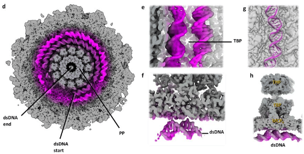
Cryo-EM resolves the structure of the archaeal dsDNA virus HFTV1 from head to tail
Daniel X. Zhang, Michail N. Isupov, Rebecca M. Davies, Sabine Schwarzer, Mathew McLaren, William S. Stuart, Vicki A.M. Gold, Hanna M. Oksanen, Tessa E.F. Quax, Bertram Daum
Systematic analysis of immune cell motility leveraging Immunemap, an open intravital microscopy atlas
Diego Ulisse Pizzagalli, Pau Carrillo-Barbera, Elisa Palladino, Kevin Ceni, Benedikt Thelen, Alain Pulfer, Enrico Moscatello, Raffaella Fiamma Cabini, Johannes Textor, Inge M. N. Wortel, The Immunemap project consortium, Rolf Krause, Santiago Fernandez Gonzalez
Sir2 is required for the quiescence-specific condensed three-dimensional chromatin structure of rDNA
Christine Cucinotta, Rachel Dell, Kris Alavattam, Toshio Tsukiyama
Spatially resolved rewiring of mitochondria-lipid droplet interactions in hepatic lipid homeostasis
Sun Woo Sophie Kang, Lauryn A Brown, Colin B Miller, Katherine M Barrows, Jihye L Golino, Constance M Cultraro, Daniel Feliciano, Mercedes B. Cornelius-Muwanuzi, Andy D Tran, Michael Kruhlak, Alexei Lobanov, Maggie Cam, Natalie Porat-Shliom
Compositional Flexibility of the ER-Mitochondria Encounter Structure
Christian Covill-Cooke, Takashi Hirashima, Shin Kawano, Joe Ganellin, Andrew Moody, Sabine N.S. van Schie, Arun T. John Peter, Chika Saito, Toshiya Endo, Benoît Kornmann
Mature tuft cell phenotypes are sequentially expressed along the intestinal crypt-villus axis following cytokine-induced tuft cell hyperplasia
Julian R. Buissant des Amorie, Max A. Betjes, Jochem Bernink, Joris H. Hageman, Maria C. Heinz, Ingrid Jordens, Tiba Vinck, Ronja M. Houtekamer, Ingrid Verlaan-Klink, Sascha R. Brunner, Dimitrios Laskaris, Jacco van Rheenen, Martijn Gloerich, Hans Clevers, Jeroen S. van Zon, Sander J. Tans, Hugo J.G. Snippert
Single cell migration along and against confined haptotactic gradients
Isabela Corina Fortunato, David B. Brückner, Steffen Grosser, Leone Rossetti, Miquel Bosch-Padrós, Jonel Trebicka, Pere Roca-Cusachs, Raimon Sunyer, Edouard Hannezo, Xavier Trepat
Serial intravital microscopy reveals temporal dynamics of autoreactive germinal centers in the spleen
Layla Pohl, Thomas R Wittenborn, Ali Shahrokhtash, Kristian Savstrup Kastberg, Cecilia Fahlquist-Hagert, Lisbeth Jensen, Donato Sardella, Alain Pulfer, Duncan S Sutherland, Santiago F Gonzalez, Ina Maria Schiessl, Soren E Degn
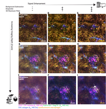
Identification of low copy synaptic glycine receptors in the mouse brain using single molecule localisation microscopy
Serena Camuso, Yana Vella, Souad Youjil Abadi, Clémence Mille, Bert Brône, Christian G Specht
Three-dimensional quantitative characterization of Coxiella burnetii infection using focused ion beam-scanning electron microscopy
Jack M. Botting, Samuel Steiner, Morven Graham, Xinran Liu, Craig R. Roy, Jun Liu
Cryogenic light microscopy with Ångstrom precision deciphers structural conformations of PIEZO1
Hisham Mazal, Alexandra Schambony, Vahid Sandoghdar
Smart 3D super-resolution microscopy reveals the architecture of the RNA scaffold in a nuclear body
Enya S. Berrevoets, Laurell F. Kessler, Ashwin Balakrishnan, Michaela Müller-McNicoll, Bernd Rieger, Sjoerd Stallinga, Mike Heilemann
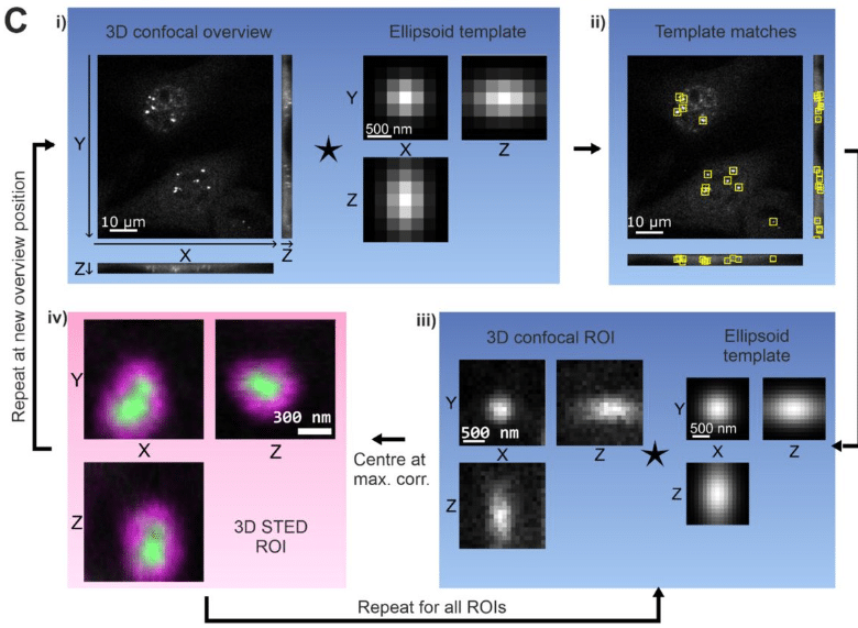
3D Reconstruction of Neuronal Allometry and Neuromuscular Projections in Asexual Planarians Using Expansion Tiling Light Sheet Microscopy
Jing Lu, Hao Xu, Dongyue Wang, Yanlu Chen, Takeshi Inoue, Liang Gao, Kai Lei
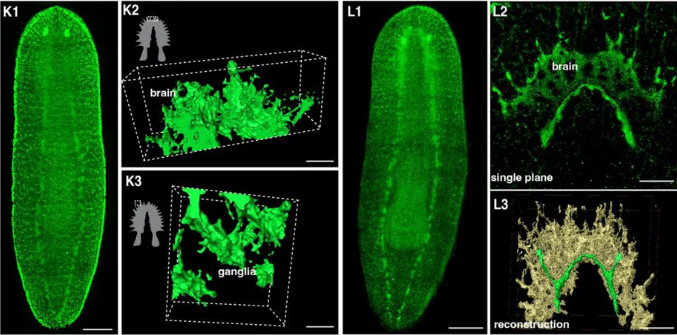
Single-Molecule Super-Resolution Imaging Reveals Formation of NS2B3 Protein Clusters on Mitochondrial Network Leading to its Fragmentation during the Onset of Dengue (Denv-2) Viral Infection
Jiby M. Varghese, Prakash Joshi, S Aravinth, Partha P. Mondal
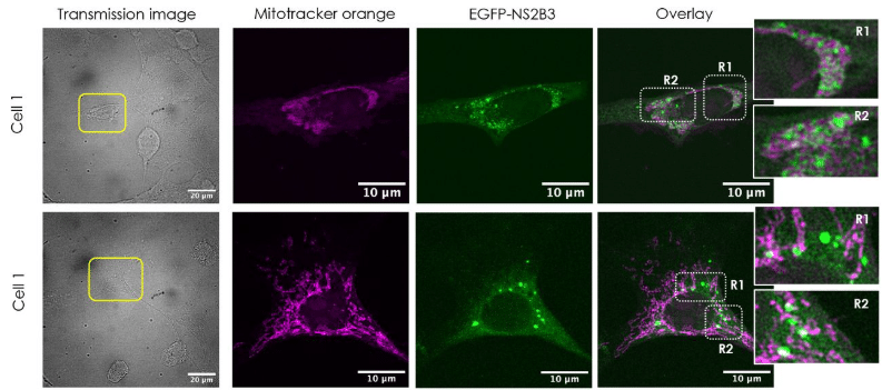
Mapping single-cell rheology of ascidian embryos in the cleavage stages using AFM
Takahiro Kotani, Tomohiro Matsuo, Megumi Yokobori, Yosuke Tsuboyama, Yuki Miyata, Yuki Fujii, Kaori Kuribayashi-Shigetomi, Takaharu Okajima
Intraflagellar Transport Selectivity Occurs with the Proximal Portion of the Trypanosome Flagellum
Aline Araujo Alves, Jamin Jung, Gaël Moneron, Humbeline Vaucelle, Cécile Fort, Johanna Buisson, Cataldo Schietroma, Philippe Bastin
Combining live cell fluorescence imaging with in situ cryo electron tomography sheds light on the septation process in Deinococcus radiodurans
L. Gaifas, J.P. Kleman, F. Lacroix, E. Schexnaydre, J. Trouve, C. Morlot, L. Sandblad, I. Gutsche, J. Timmins
Pearling Drives Mitochondrial DNA Nucleoid Distribution
Juan C. Landoni, Matthew D. Lycas, Josefa Macuada, Roméo Jaccard, Christopher J. Obara, Andrew S. Moore, Sheda Ben Nejma, David Hoffman, Jennifer Lippincott-Schwartz, Wallace Marshall, Gabriel Sturm, Suliana Manley
Direct Intercellular Vesicle Exchange between Adjacent Cells
Tomohiro Minakawa, Tatsuya Katsuno, Keiko Okamoto-Furuta, Ryoko Ando, Fumiyoshi Ishidate, Takahiro K. Fujiwara, Dooseon Cho, Sho Takehana, Yoshikatsu Sato, Yasuhiko Tabata, Atsushi Miyawaki, Jun K. Yamashita
Dynamic remodeling of centrioles and the microtubule cytoskeleton in the lifecycle of chytrid fungi
Alexandra F. Long, Krishnakumar Vasudevan, Andrew J.M. Swafford, Claire M. Venard, Jason E. Stajich, Lillian K. Fritz-Laylin, Jessica L. Feldman, Tim Stearns
DNAJC13 localization to endosomes is opposed by its J domain and its disordered C- terminal tail
Hayden Adoff, Brandon Novy, Emily Holland, Braden T Lobingier
The Chromosome Periphery is an Essential Compartment of Oocyte Chromosomes
Eva L Simpson, Ben Wetherall, Aleksandra Byrska, Liam P Cheeseman, Tania Mendonca, Xiaomeng Xing, Alison J Beckett, Helder Maiato, Alexandra Sarginson, Ian A Prior, Geraldine M Hartshorne, Andrew D McAinsh, Suzanne Madgwick, Daniel G. Booth
Scale invariance of mechanical properties in the developing mammalian retina
Elijah Robinson Shelton, Michael Frischmann, Achim Theo Brinkop, Rebecca Marie James, Friedhelm Serwane
Cell-sheet shape transformation by internally-driven, oriented forces
Junrou Huang, Juan Chen, Yimin Luo
Actin and vimentin jointly control cell viscoelasticity and compression stiffening
James P. Conboy, Mathilde G. Lettinga, Pouyan E. Boukany, Fred C. MacKintosh, Gijsje H. Koenderink
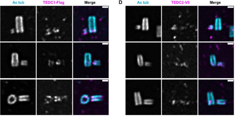
A delta-tubulin/epsilon-tubulin/Ted protein complex is required for centriole architecture
Rachel Pudlowski, Lingyi Xu, Ljiljana Milenkovic, Chandan Kumar, Katherine Hemsworth, Zayd Aqrabawi, Tim Stearns, Jennifer T. Wang
Nucleus softens during herpesvirus infection
Aapo Tervonen, Simon Leclerc, Visa Ruokolainen, Katie Tieu, Sébastien Lyonnais, Henri Niskanen, Jian-Hua Chen, Alka Gupta, Minna U Kaikkonen, Carolyn A Larabell, Delphine Muriaux, Salla Mattola, Daniel E Conway, Teemu O Ihalainen, Vesa Aho, Maija Vihinen-Ranta


 (No Ratings Yet)