Imaging with… INDICASAT AIP Advance Microscopy and Bioimaging Core Facility
Posted by FocalPlane, on 5 December 2025
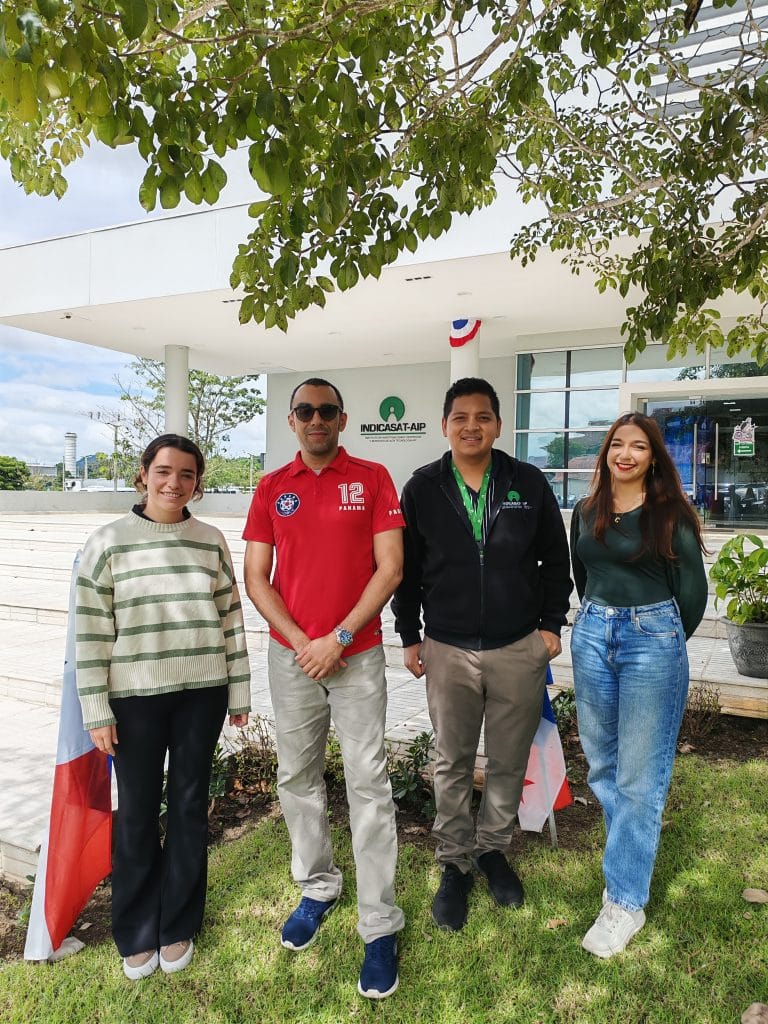
In our ‘Imaging with…’ blog post, we meet the team at Nucleo de Microscopia Avanzada y Bioimagenes, Instituto de Investigaciones Científicas y Servicios de Alta Tecnología de Panamá.
Staff role call
Armando Castillo PhD, Core Facility Coordinator
Expertise: Neurophysiology, Microscopy
Most likely to be found making coffee for everyone, eating desserts and helping others with their research processes. Also writing emails, proposals and papers, or learning about new microscopes and techniques.
Carolina Alvarado, Confocal Microscope Techician
Expertise: Advanced confocal microscopy techniques applied to neuroscience, parasitology, and cell biology research; Z-stack, stitching, DIC, and other imaging techniques for nervous tissue, cell cultures, and small organisms, ensuring high-resolution imaging while preserving sample integrity. Contributions include studies on nuclear translocation, parasite analysis (Leishmania, Cryptosporidium), viral infections, rat brain tissue visualization, neuronal structures, and cell samples on titanium, among others.
Most likely to be foundreading fantasy books, eating sweets with her coworkers or taking amazing and colourful pictures in the lab.
Andrea Berraondo, Image analysis expert
Expertise: Machine learning for identification of cells, tissues and biological structures.
Most likely to be found programming on the laptop.
Kevin Amaya, Scanning Electron Microscope Technician
Expertise: Operating the SEM across all three vacuum modes, using the cooling and heating stages, performing EDS analysis, and working with secondary electrons, backscattered electrons, and STEM imaging. Additional expertise includes acquiring images of cells adhered to biomaterials, semiconductor nanostructures, nanoparticles, insects, parasites, fossils, and other diverse sample types.
Most likely to be found listening to music, half-frozen in the lab, taking photos of some random bug.
Microscope role call:
The Quattro S from ThermoFisher Scientific is an environmental field-emission scanning electron microscope equipped with three vacuum modes—high, low, and environmental—along with precise control of temperature, pressure, and humidity inside the chamber. This versatility makes it an ideal tool for analyzing a wide range of samples in materials science and biological research. The system offers resolutions of 1.08 nm in high vacuum, 3.16 nm in low vacuum, and 0.75 nm in STEM mode, with a magnification range from 6× to 2,500,000× and an acceleration voltage between 20 eV and 30 keV. It is equipped with multiple detectors—including ETD, LVD, GSED, ABS/CBS, STEM, and EDS—and features a multi-purpose stage capable of handling various samples, with cooling and heating options to accommodate diverse experimental needs.
The Olympus FV3000 confocal microscope is a laser-scanning confocal system designed for biomedical research, life-science applications, and advanced imaging analysis. It is a highly versatile instrument that delivers exceptional resolution, sensitivity, and acquisition speed for both live-cell and fixed-sample studies. The system includes 405 nm, 488 nm, 561 nm, and 640 nm lasers, paired with objective lenses of 4×, 10×, 20×, 40×, 60×, and 100×, enabling detailed visualization from cellular to subcellular structures. It supports multiple acquisition modes—including 2D, 3D, Z-stack, time-lapse, FRAP, and FRET—and uses the FluoView software platform for advanced image processing and analysis.
Who can access the facility?
The institute provides microscopy services to both internal and external users — mainly researchers and their thesis students, interns, or anyone interested in observing their samples. We also occasionally host visits from students or other research centers to showcase what we’ve been able to achieve with our equipment.
Pet peeve (something that users do that is annoying): When people show up late or without warning… and especially when they have no idea what they want to look at.
Favourite microscope: It’s a tie! The FV3000, because we get beautifully colorful, very artistic samples. And the Quattro S (SEM), because you can get insanely high-resolution images even from biological samples suspended in water.
Favourite thing to image: Virus-infected cells stained with red and blue dyes to create a 3D effect, as well as cells interacting with or adhering to biomaterials.
Best bit of advice (that you give or have been given)
Don’t give up, science can be slow but it will work out! — keep searching, keep learning, read every paper you need to..
If money was no object, we would buy… a cryo-TEM and a multiphoton microscope.
Can you give us some examples of recent papers that were published with your assistance?
Jaén, J.A., Coronado, M., Chung, E. et al. Structural and electrochemical characterization of tetragonal copper ferrite nanoparticles. Interactions 245, 4 (2024). https://doi.org/10.1007/s10751-024-01848-7
https://www.researchgate.net/publication/377413609_Structural_and_electrochemical_characterization_of_tetragonal_copper_ferrite_nanoparticles
Freire, A., Chung, E., Mendoza, I. et al. Green synthesis of iron oxide nanoparticles using Caesalpinia coriaria (Jacq.) Willd. fruits extract. Hyperfine Interact 244, 6 (2023). https://doi.org/10.1007/s10751-023-01817-6.
https://www.researchgate.net/publication/367388671_Green_synthesis_of_iron_oxide_nanoparticles_using_Caesalpinia_coriaria_Jacq_Willd_fruits_extract
How should users acknowledge the facility and why is it important?
- “The co-authors would like to acknowledge the INDICASAT AIP Microscopy and Bioimaging Core Facility for the use of the FV-3000 confocal microscope.
- The co-authors would like to acknowledge the INDICASAT AIP Microscopy and Bioimaging Core Facility for the use of the Thermo Quattro S eSEM.
Or
- “The co-authors would like to acknowledge INDICASAT AIP Microscopy and Bioimaging Core Facility for use of the FV-3000 Confocal Microscope and staff X for helpful discussions on sample preparation.”
 (No Ratings Yet)
Get involved
Create an account or log in to post your story on FocalPlane.
More posts like this

Filter by
- NewsApply
- DiscussionsApply
- How toApply
- ToolsApply
- Case studiesApply
- InterviewsApply
- JobsApply
- EducationApply
- Blog seriesApply
- Volume EMApply
- Latin American Micro..scopistsApply
- Bio-image Analysis w..ith NapariApply
- Imaging with…Apply
- Towards Global Acces..sApply
- Latin America Bioima..gingApply
- From Zero to Qupath ..HeroApply
- Asian Microscopists ..and Cell BiologistsApply
- AIC at HHMI JaneliaApply
- Deep Learning for Bi..o-image analysisApply
- GloBIAS – updates fr..om the communityApply
- WAMBIAN: West Africa.. in FocusApply
- Highlights from Euro..-BioImagingApply
- LSFM seriesApply
- DIY MicroscopyApply
- View all