Imaging spotlight: Revealing mouse brain ultrastructure using non-destructive X-ray tomography
Posted by FocalPlane, on 13 January 2026
In this paper highlight, Carles Bosch, Ana Diaz, Adrian A. Wanner and Andreas T. Schaefer (on behalf of all authors) describe how they use nondestructive X-ray tomography to map mouse brain tissue ultrastructure .
What’s the technique and what’s new about your method?
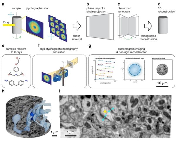
Tracing all cells in a tissue volume is key to gathering mechanistic understanding on the function of that tissue – be that when studying neuronal circuits in the brain, cancerous and healthy neighbouring cells in a tumour, or host and pathogen relationships in an infected tissue. X-rays have the potential to boost the capacity to obtain those maps, but their applicability to nanoimaging in biological tissues has been hampered by sample degradation: the X-rays needed to obtain the necessary resolution would disintegrate the sample long before obtaining the dataset. We assembled solutions along the process (in sample preparation, in imaging and in data reconstruction) to lift this limitation, and recovered sufficient resolution to detect synapses in mouse brain tissue.
How has your method been used so far?
Ptychographic tomography has been used in imaging nanometer details in volumes relevant for materials science – from cement to computer chips. In the latter, it recently demonstrated its capacity to resolve circuit ports of 4nm in size. Our study demonstrates its applicability to resolve details finer than 40nm in brain tissue, and signals avenues to further improve in both resolution and imaging speed.
What’s next in the application of this technique?
Imaging ultrastructure in 3D with X-rays comes with the advantage of not having to slice the tissue in lengthy series of ultrathin sections. This brings multiple advantages: not only samples are not lost during imaging, but most importantly this lifts the rate-limiting step in tissue nanoimaging. Our insights, applied to higher-throughput imaging X-ray phase contrast regimes at 4th-generation synchrotron sources, can boost the throughput of tissue nanoimaging, making volumes of a cubic millimetre and beyond more viable to obtain in a routine manner.
What are the basic requirements for a researcher to image their own samples?
- Samples need to be prepared in a way they can withstand high fluxes of X-rays through them. We introduce the use of a resin initially developed for the aerospace industry, TGPAP-DDM, that allows irradiating tissues in a way that beyond 1010 Gy are absorbed in the sample.
- Imaging algorithms should be resilient to mild sample deformations during the acquisition. We implemented a subtomogram acquisition and non-rigid reconstruction process (Odstrcil, et al.), that can counteract those effects to some extent.
- Finally, nanoimaging X-ray phase contrast beamlines. These are available at multiple synchrotrons worldwide, openly accessible to any scientist. Scientists based in any country only need to apply with a competitive proposal to run an experiment in any synchrotron. Most synchrotrons open calls twice a year, and not only provide the awarded beamtime at no additional cost, but also often subsidize travel-associated costs.
What are the prospects for further development?
- Resolution: While we demonstrated that X-rays can resolve tissue ultrastructure at 40nm detail, there is great expectation for improving that resolution below the 20nm mark. At that accuracy, all plasma membranes will be resolved in 3D, enabling full reconstruction of cells in tissues – and therefore connectomics studies using X-rays.
- Volume: We present our results in brain samples that were prepared to match the best performance of ptychographic tomography, but neuronal circuits in mammalian brains (and other biological systems in tissues) typically occupy larger volumes. Reaching the cubic millimetre milestone might be better suited incorporating other versions of X-ray phase contrast tomography, such as full-field holographic regimes.
- Throughput: Volumes of interest at connectomics resolution should be acquired within the length of a typical beamtime – less than a week. 4th generation synchrotron sources provide sufficient light flux and beam coherence to reach this goal (Du, et al.). Some synchrotrons, like ALBA and ESRF, have already committed to constructing nanoimaging beamlines dedicated to biological tissues in their 4th generation rings, and other light sources are following suit.
Where can people find more information?
You can check our paper here: Bosch, C., Aidukas, T., Holler, M., Pacureanu, A., Müller, E., Peddie, C. J., Zhang, Y., Cook, P., Collinson, L., Bunk, O., Menzel, A., Guizar-Sicairos, M., Aeppli, G., Diaz, A., Wanner, A. A. & Schaefer, A. T. Nondestructive X-ray tomography of brain tissue ultrastructure. Nature methods, doi:10.1038/s41592-025-02891-0 (2025).
You can also explore the datasets we analysed in this study (100+ 3D browser-accessible datasets) and other details (like code behind key figures) in the public repository ptychoStainedTissue.
Bosch, C., Aidukas, T., Holler, M., Pacureanu, A., Müller, E., Peddie, C. J., Zhang, Y., Cook, P., Collinson, L., Bunk, O., Menzel, A., Guizar-Sicairos, M., Aeppli, G., Diaz, A., Wanner, A. A. & Schaefer, A. T. in Zenodo Non-destructive X-ray tomography of brain tissue ultrastructure. Supporting code ptychoStainedTissue. [Software]. 10.5281/zenodo.17233854. Code version released alongside manuscript publication. (Zenodo, 2025).
 (No Ratings Yet)
Get involved
Create an account or log in to post your story on FocalPlane.
More posts like this


Filter by
- NewsApply
- DiscussionsApply
- How toApply
- ToolsApply
- Case studiesApply
- InterviewsApply
- JobsApply
- EducationApply
- Blog seriesApply
- From Zero to Qupath ..HeroApply
- Asian Microscopists ..and Cell BiologistsApply
- AIC at HHMI JaneliaApply
- Deep Learning for Bi..o-image analysisApply
- GloBIAS – updates fr..om the communityApply
- WAMBIAN: West Africa.. in FocusApply
- Volume EMApply
- Latin American Micro..scopistsApply
- Bio-image Analysis w..ith NapariApply
- Imaging with…Apply
- Towards Global Acces..sApply
- Latin America Bioima..gingApply
- Highlights from Euro..-BioImagingApply
- LSFM seriesApply
- DIY MicroscopyApply
- View all