Imaging with… Unidad de Microscopía, Universidad Mayor, Chile
Posted by FocalPlane, on 2 February 2026
In our ‘Imaging with…’ blog post, we meet the team at Unidad de Microscopía, Universidad Mayor, Chile.
Staff role call
Luz María Fuentealba, Facility Manager
Expertise:Fluorescence Microscopy
Most likely to be found roaming between microscopes or chasing down users.
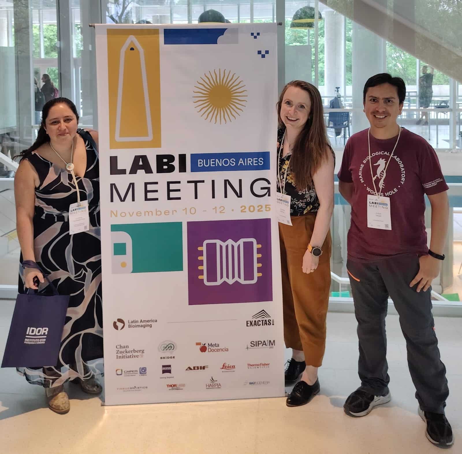
Aníbal Vargas, Light-sheet Specialist
Expertise:Light-sheet Microscopy
Most likely to be in the light-sheet room or looking for a good spot to work on the computer.
Charlotte Buckley, Facility Director
Expertise:Live imaging on pretty much every microscope
Most likely to be found in her office writing grants to secure funding but wishing she was imaging.
Microscope role call
Zeiss LS7 Light-sheet microscope (in-person and remote imaging are available, but you can also send your samples to be imaged by us), Flamingo light-sheet, Leica DMi8, Leica SP8 Confocal microscope, Zeiss Apotome 3, CytoFLEX, various image analysis stations
Pet peeve (something that users do that is annoying): Currently, our top pet peeve is users arriving late to their sessions. Obviously, there are lots of different reasons for this, but as facility staff we’re already so busy trying to keep many plates spinning, so having to chase people down to know whether they will use the microscope always feels like a waste of time.
Favourite microscope: The light-sheet is so fast! It’s also fantastic for playing with different types of samples and allows us to get creative with how we mount the samples. Having said that, the confocal is also one of our favourites because is usually the most reliable, easy to use, and you can get a nice image from almost everything.
Favourite thing to image: Bichos! As part of an outreach initiative in Chile called MOLA (Museos, Observatorios y Laboratorios Abiertos), we invited 12 and 13 year-old school children to learn about microscopy, and the types of experiments that we run in the lab. As part of this, we took them into the gardens surrounding the University to find exciting bugs and plants to image. They loved it, and so did we! As everyone who is reading this probably knows, everything looks amazing under a microscope, especially insect eyes!
Best bit of advice (that you give or have been given): “When in doubt, ask”. There always tends be someone who’s had a similar problem, and it’s so much faster for us to tackle the problem together.
If money was no object, we would buy… Talking about this between the team, we realised that our lists are quite long… As well as a mesoSPIM to image larger, fully clarified samples, and a Dragonfly with TIRF and single molecule capabilities to map intracellular trafficking processes, we realised that what we’d love to have is a proper data server. Whilst using the light-sheet, we acquire increasingly large datasets, both from timelapse imaging sessions of developmental processes, and from whole clarified organs. Without a server, it’s extremely difficult to store and analyse the data to its fullest extent, so having one would not only help with this but would also allow us to collaborate with the community more effectively.
Who can access the facility?
Anyone! We have different pricing structures for internal and external users from academic institutions, and for industry partners. Many of our users are from biomedicine and study cells, tissue preparations, zebrafish and Drosophila. However, we also work with samples from around Chile, with sponges from Antarctica to amphipods from the Pacific.
Here in UM2 we have the LiSIUM initiative, whose goal is to democratize the cutting-edge light-sheet technology that we have here at Universidad Mayor. As the only commercial light-sheet available in Chile, and the first system in South America, we are passionate about spreading our experiences through hands-on training, online seminars and annual workshops on different areas of light-sheet imaging and analysis. We have national and international users who will send their samples to us for imaging, and others who will come with their samples to use the system.
Can you give us some examples of recent papers that were published with your assistance?
Confocal and first paper from our Centre using our light-sheet: A gal4 insertion in the rx3 locus as a tool for visualization and manipulation of eye fated cells in zebrafish
How should users acknowledge the facility and why is it important?
We follow the Global BioImaging and RMS initiative which aims to provide a framework for appropriate acknowledgement of facility work in scientific publications.
For us, it’s incredibly important that the work performed in the facility is acknowledged. In general, the skills of facility managers and technicians have been undervalued, and appropriate acknowledgement is one of the few ways for us to gain visibility and show our worth to the University and the wider community.
We also depend on funding from the Chilean Government for equipment, and in order for us to grow and maintain our services, we need to show that we are fully integrated into the research community. It’s important to show that researchers depend on our skills and expertise to bring in future grant funding.
 (1 votes, average: 1.00 out of 1)
One thought on “Imaging with… Unidad de Microscopía, Universidad Mayor, Chile”
Leave a Reply
Get involved
Create an account or log in to post your story on FocalPlane.
More posts like this


Filter by
- NewsApply
- DiscussionsApply
- How toApply
- ToolsApply
- Case studiesApply
- InterviewsApply
- JobsApply
- EducationApply
- Blog seriesApply
- Volume EMApply
- Latin American Micro..scopistsApply
- Bio-image Analysis w..ith NapariApply
- Imaging with…Apply
- Towards Global Acces..sApply
- Latin America Bioima..gingApply
- From Zero to Qupath ..HeroApply
- Asian Microscopists ..and Cell BiologistsApply
- AIC at HHMI JaneliaApply
- Deep Learning for Bi..o-image analysisApply
- GloBIAS – updates fr..om the communityApply
- Highlights from Euro..-BioImagingApply
- LSFM seriesApply
- DIY MicroscopyApply
- View all
It is great to see the UM2 team featured on FocalPlane. This recognition goes beyond infrastructure; it celebrates a profound commitment to the democratization of science. And I particularly appreciate the focus on the expertise of the facility staff.
As the article suggests, the transition from raw photons to meaningful data relies entirely on their guidance. Moreover, their outreach work is a beautiful reminder that cutting-edge microscopy should always be rooted in a sense of wonder for the natural world. Congratulations to the team for putting Chilean bioimaging at the center of the global focal plane!