

Microscopy preprints: bioimage analysis
Posted by FocalPlane, on 12 June 2026
Here is a curated selection of preprints published or updated recently. In this post we focus specifically on bioimage analysis and data management.
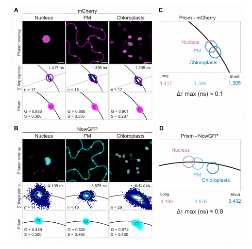
Fluorescence Lifetime Imaging in Plants: Practical guidelines for multiplexing, label-free imaging and data analysis
Beatrice Lace Dr., Pascal Krohn Dr., Morgane Batzenschlager Dr., Vanessa Fiess, David Molina Dr., Laura Ragni Dr., Thomas Ott Prof.
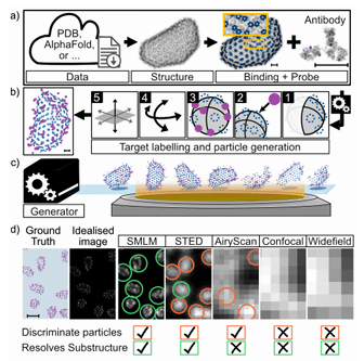
VLab4Mic: prediction of structural resolvability in super-resolution microscopy
Damián Martínez, Bruno M. Saraiva, Tayla Shakespeare, Mark Bates, Dylan M. Owen, Christophe Leterrier, Mario Del Rosario, Ricardo Henriques
Vermeer: Autoregressive generative modeling of microscopy predicts protein localization
Sandeep Kambhampati, Eric Zimmermann, Emre Hayir, Kevin K. Yang, Fei Chen, Alex X. Lu
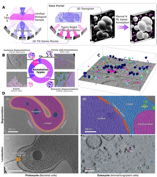
POPSICLE: Benchmark Datasets for Segmentation and Localization in CryoET
Jonathan Schwartz, Utz Heinrich Ermel, C. Braxton Owens, Zhuowen Zhao, Ariana Peck, Gus L.W. Hart, Grant J. Jensen, Bridget Carragher, Dari Kimanius
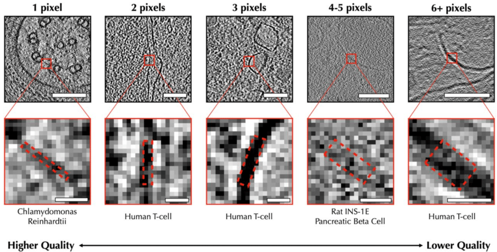
TomoScore: A Neural Network Approach for Quality Assessment of Cellular cryoET
Xuqian Tan, Xueting Zhou, Ethan Boniuk, Anisha Abraham, Zhili Yu, Valerie Dalton, Steven J. Ludtke, Zhao Wang
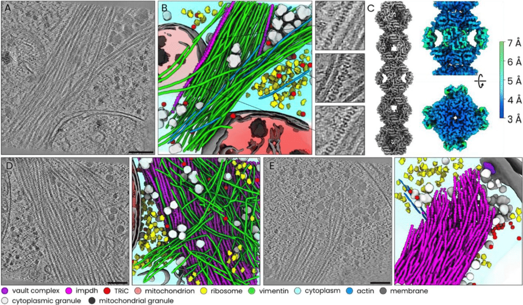
Easymode: general pretrained networks for cellular cryo-ET enable flexible approaches to subtomogram averaging
Mart G. F. So-Last, Alister Burt, Thomas Hale, Matteo Allegretti
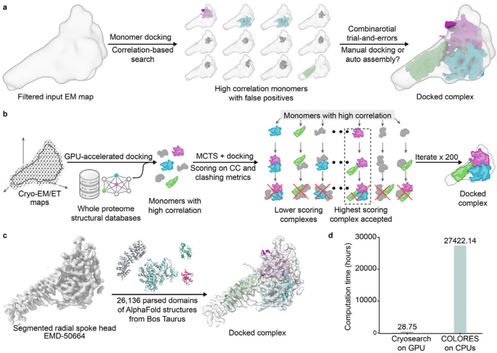
Automated assembly of protein complexes from cryo-EM maps with structure-informed Monte Carlo Tree Search
Rohit Dilip, Songrong Jeff Qu, Zhen Chen, David Van Valen
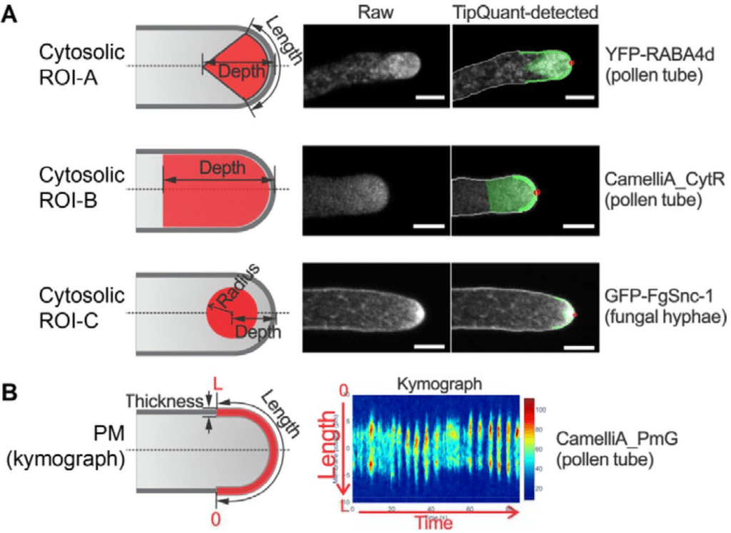
TipQuant: A robust algorithm for quantitative analysis of spatiotemporally dynamic activities in tip-growing cells
Jingzhe Guo, Julian Le Gouic, Rémi Rosenthal, Ailing Zou, Xiang Zhou, Nicolas Brunel, Zhenbiao Yang, Xinping Cui
Celldetective: an AI-enhanced image analysis tool for unraveling dynamic cell interactions
Rémy Torro, Beatriz Díaz-Bello, Dalia El Arawi, Ksenija Dervanova, Lorna Ammer, Florian Dupuy, Patrick Chames, Kheya Sengupta, Laurent Limozin
A Decade of Deep Learning-based Biomedical Image Segmentation
Suhao Yu, Haojin Wang, Ningsen Wang, Sicheng Chen, Juncheng Wu, Zhenlong Yuan, Tianhao Qi, Zongwei Zhou, Fei Xia, Jun Ma, Yuyin Zhou
OMIO: A policy-driven Python library for reproducible microscopy image I/O
Fabrizio Musacchio, Henrike Antony, Sophie Crux, Falko Fuhrmann, Nala Gockel, Denise Marie Hoffmann, Dilek Mercan, Felix Christopher Nebeling, Martin Fuhrmann
ATI_Box: A Simple tool for convolutional neural network-based image semantic segmentation
Tomasz Przygodzki
Supervised Deep Learning for Efficient Cryo-EM Image Alignment in Drug Discovery with cryoPARES
Ruben Sanchez-Garcia, Alex Berndt, Amir Apelbaum, Judith Reeks, Pamela A Williams, Carl Poelking, Charlotte M Deane, Michael Saur
TetraFuse: A Synergistic Four-Dimensional Dynamic Fusion Framework for Efficient and Robust Medical Image Classification
Yufei Gao, Jiaqi Li, Jing Xu, Qing Li, Ziyu Li, Guohua Zhao, Yucheng Shi, Xia Wu, Yameng Zhang
SortIT — A Tool For Assessing Observer Variability And Creating Ground Truth Image Classification Datasets
Wataru Uegami, Tom Bisson, Ethan N. Okoshi, Frederico Gaia Costa da Silva, Jijgee Munkhdelger, Kris Lami, Norman Zerbe, Andrey Bychkov, Junya Fukuoka
Quantifying the spatio-temporal image degradation under motion blur in fluorescence microscopy
Serafim Korovin, Kutay Ugurlu, Dylan Kalisvaart, Manon Kok, Rainer Heintzmann, Kirti Prakash, Carlas S. Smith
Mycol: A user-friendly app for automating analysis of microscopy images
Samuel Alan Bradley, Henry Webel, Giovanni Schiesaro, Marcel Skumantz, Olga Novillo-Sanjuan, Alexandra Panagou, Rafael Lucena-Marín, Emil D. Jensen, Antonio Di Pietro, Carlos G. Acevedo-Rocha
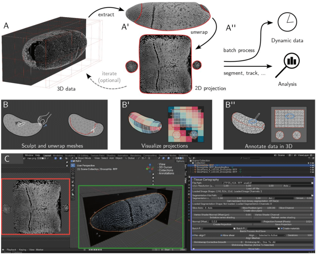
Blender tissue cartography: an intuitive tool for the analysis of dynamic 3D microscopy data
Nikolas Claussen, Cécile Regis, Susan Wopat, Matthew F. Lefebvre, Sebastian Streichan
Volumetric Denoising: An Approach to Boost Acquisition and Downstream Analysis of Volume Electron Microscopy
Bohao Chen, Fangfang Wang, Haoyu Wang, Yanchao Zhang, Zhuangzhuang Zhao, Haoran Chen, Hua Han, Xi Chen, Yunfeng Hua
3DBrainOne: an integrated end-to-end platform for 3D histological analysis of whole mouse brains
Daye Kim, Young-Gyun Park
Top Model Decision Tree: Selecting Segmentation Models for Reliable Quantitative Analysis in Low- and Ultralow-Dose CryoEM
Lynnicia N. Massenburg, Sita S. Madugula, Spenser R. Brown, Amber N. Bible, Chanda R. Harris, Lance X. Zhang, Kiara Parker, Scott T. Retterer, Jennifer L. Morrell-Falvey, Rama K. Vasudevan, Alexis N. Williams
Instant Prior-Free Resolution Enhancement for Cross-Modality Microscopy
Haohong Gan, Shiyi Peng, Hailian Hu, Xuan You, Yabo Guo, Ruiyang Guo, Ziliang Chen, Jun Qian
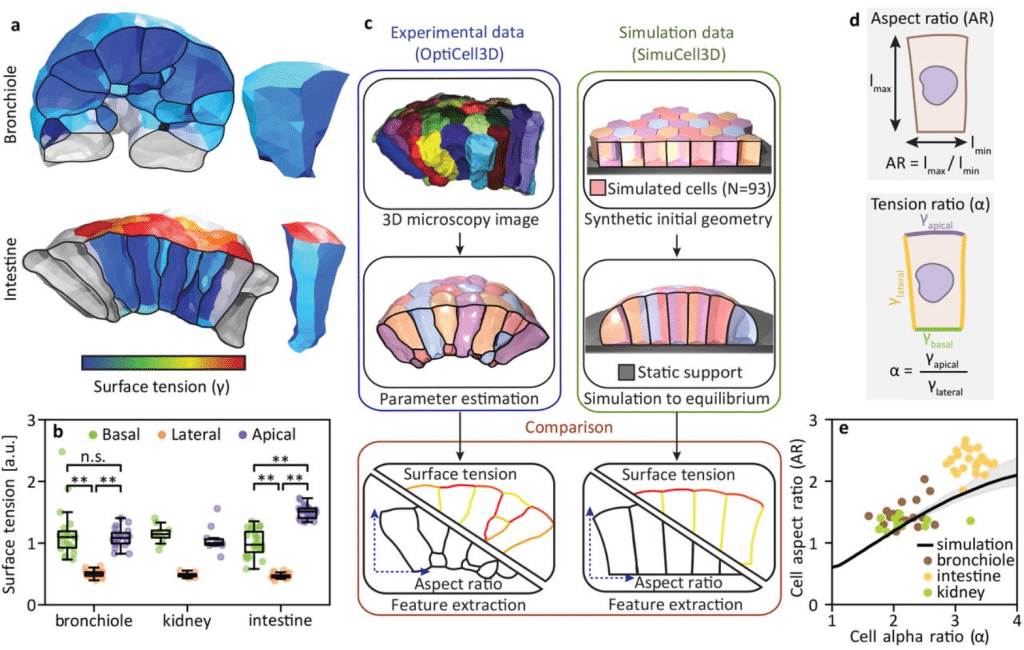
OptiCell3D: Precise inference of mechanical cell properties from microscopy imaging
Steve Runser, Kevin A Yamauchi, Marius Almanstötter, Franziska L Lampart, Federico Carrara, Lisa Conrad, Laura Schaumann, Roman Vetter, Dagmar Iber
BaSiCPy: Scalable and Robust Shading Correction for Optical Microscopy Images
Yu Liu, Yohsuke T. Fukai, Santiago Cano-Muniz, Victor Perez, Mihail Todorov, Germán Camargo Ortega, Timothy Morello, Alexander Jovanovic, Dirk Loeffler, Johannes C. Paetzold, Xun Xu, Phillipp Paulischka, Lorenz Lamm, Nan Ma, Ali Erturk, Timm Schroeder, Lucas Boeck, Denis Schapiro, Nicholas Schaub, Carsten Marr, Tingying Peng
Self-supervised Internal Learning Enhances Isotropic Resolution for Three-dimensional Fluorescence Microscopy
Mingzhe Wei, Pengcheng Xu, Junyu Liu, Xuesong Li, Xuhui Feng, Jun Zhu, Renwei Dong, Hengjia Ran, Wentao Zhu, Yubing Han, Yue Li, Min Guo, Huafeng Liu
CiliAI: Automated segmentation and compartment-specific fluorescence quantification of primary cilia in confocal microscopy images
Eliza Karapetian, Christoph Gerhardt, Emrah Nazif, Thorsten Pfirrmann
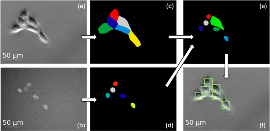
LFCT: A Benchmark Dataset for Low-Frame-Rate Cell Tracking in Long-Term Live-Cell Microscopy
Mina Gachloo, Tirthankar Biswas, Xiaoming Lu, Caroline M. Greene, Chandler K. Hargett, Katelyn R. Simancik, Marc R. Birtwistle, Federico Iuricich
Topology-aware reconstruction of cellular state landscapes from microscopy using self-supervised learning
Elisa Messori, Doaa Taha, Lisa Fournier, Anna Foix Romero, Virginie Uhlmann, Pascal Frossard, Cédric Vincent-Cuaz, Rickie Patani, Raphaëlle Luisier
SPIFEE — A pipeline for analyzing traces of live-cell fluorescence microscopy data
Colin Hogendorn, Ingrid R. Aragon, Samuel Dallon, Eric Batchelor
Universal approach to wave-optical calculations of point spread functions in microscopy (and beyond)
Ivan Gligonov, Lars Loetgering, Francisco Tenopala-Carmona, Chia-Lung Hsieh, Ingo Gregor, Jörg Enderlein
Automated Multimodal Correlative Registration for Organelle-Specific Molecular Imaging
Chixiang Lu, Kaiqiang Zhao, Di Cui, Gu Chen, Qian Yang, Hui Yang, Murong Zhao, Kaiyun Song, Mehran Nikan, Zhijie Li, Shanchao Zhao, Jinpeng Cen, Xincheng Qiu, Stephen G. Young, C. Frank Bennett, Punit Seth, Kai Chen, Xiaojuan Qi, Haibo Jiang
A general method for bootstrapping dense 3D segmentations from sparse 2D annotations
Vijay Venu Thiyagarajan, Arlo Sheridan, Kristen M. Harris, Uri Manor
Agentic-J: An AI Agent for Biological Microscopy Image Analysis
Lukas Johanns, Marilin Moor, Davide Panzeri, Yu Zhou, Xinyi Chen, Nora F. K. Pauly, Zixuan Pan, Matthias Gunzer, Andreas Müller, Yiyu Shi, Hedi Peterson, Jianxu Chen
MicroscopyMatching: Towards a Ready-to-use Framework for Microscopy Image Analysis in Diverse Conditions
Xiaofei Hui, Haoxuan Qu, Hossein Rahmani, Shuohong Wang, Jeff W. Lichtman, Jun Liu