Microscopy preprints – new tools and techniques in imaging
Posted by FocalPlane, on 26 June 2026
Here is a curated selection of the latest preprints on new tools and techniques in imaging. Let us know if we are missing any recent preprints that are on your reading list!
Thousandfold Expansion Microscopy
Helena Hu, Donatus Krah, Antonios Ntolkeras, Sushovan Chanda, Alina Heimbrodt, Milton Mondal, Jonas Altendorf, Bowen Jing, Bonnie Berger, Ali H. Shaib, Silvio O. Rizzoli, Edward S. Boyden
OptiFoot: a method for recording protein footprints on DNA for microscopy and sequence analysis
Rūta Gerasimaitė, Jonas Bucevičius, Dovilė Bubnytė, Tanja Koenen, Gražvydas Lukinavičius
Electroporation-mediated delivery of protein biosensors for metabolic imaging in differentiated myotubes
Aki Kawamura, Cong Quang Vu, Nahoko Shimizu, Tsubasa Shibaguchi, Kazumi Masuda, Satoshi Arai
SurpHer: a genetically encoded ratiometric sensor for dynamic extracellular pH imaging
Sofie Cens Holste, Leïla Dos Santos, Mahdi Rezayati Charan, Oline Nyhegn-Eriksen, Roxane Crouigneau, Birthe B. Kragelund, Rodolphe Marie, Albin Sandelin, Jamie Yam Auxillos, Stine Falsig Pedersen
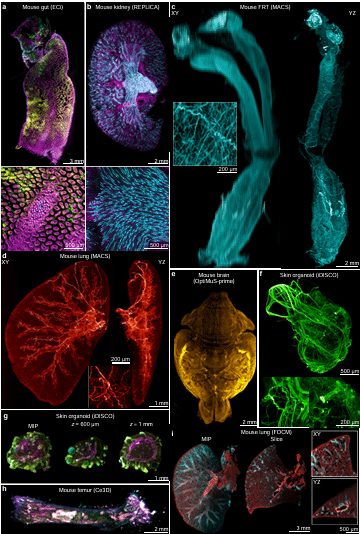
Index-agnostic oblique plane light sheet microscopy of centimetre-scale cleared tissues at subcellular resolution
Jacob R Lamb, Miguel Cardoso Mestre, Ksena Fenwyn Longrin, Pratiksha Bhat, Molly Stevenson, Anna D Y Rhodes, Joanna Gosieniecka, Leah C Redmond, Claire A Higgins, Noe Rodríguez-Rodrígues, Madeline A Lancaster, James D Manton
Revealing the spatiotemporal dynamics of methionine metabolism with a genetically encoded single-fluorophore biosensor
Katarina A. Cohen, Arnav Jhawar, Gilberto Garcia, Naiara Goni, Megan Chen, Athena Alcala, Ryo Higuchi-Sanabria, Danielle L. Schmitt
Low-Energy Polishing Facilitates Breaking the Resolution Barrier of In Situ Cryo-EM
Chunling Wu, Qi Yang, Xiaodong Su, Mei Li, Xinzheng Zhang
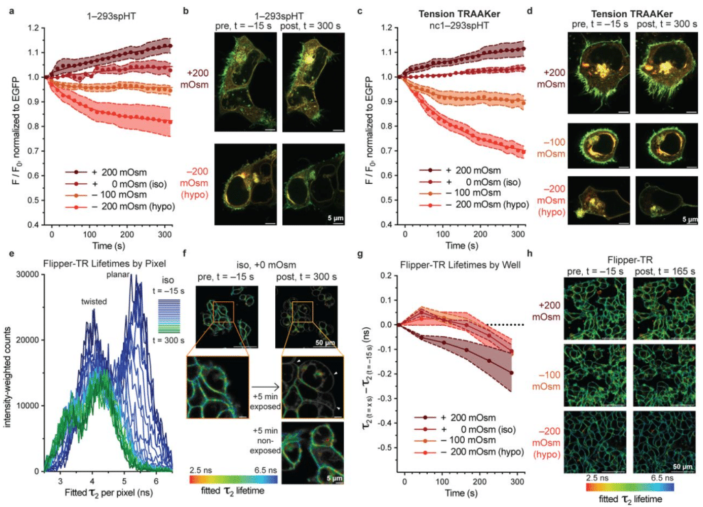
Tension TRAAKer: a chemigenetic fluorescent membrane tension reporter
Anna V. Elleman, Nels Gerstner, Benjamin E. Smith, Evan W. Miller, Richard H. Kramer, Stephen G. Brohawn
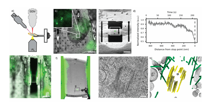
High Accuracy Fluorescence Guided Focused Ion Beam Milling
Davis Perez, Sophia Betzler, Patrick Cleeve, Carmela Villegas, Cali Antolini, Sven Klumpe, Jonathan Schwartz, John M. Mitchels, Shu-Hsien Sheu, Peter D. Dahlberg, Bridget Carragher, David A. Agard, Julia Peukes, Garrett Greenan
Near-Infrared Turn-On Fluorogenic Probe for Versatile Detection of Inorganic Polyphosphates
Kenji Torii, Rūta Gerasimaitė, Gražvydas Lukinavičius
Correlative X-ray imaging and fluorescence microscopy
Mangalika Sinha, Boram Yu, Rita Mendes da Silva, Ulrike Rölleke, Peter Luley, Malte Tiburcy, Wolfram-Hubertus Zimmermann, Manfred Burghammer, Sarah Köster
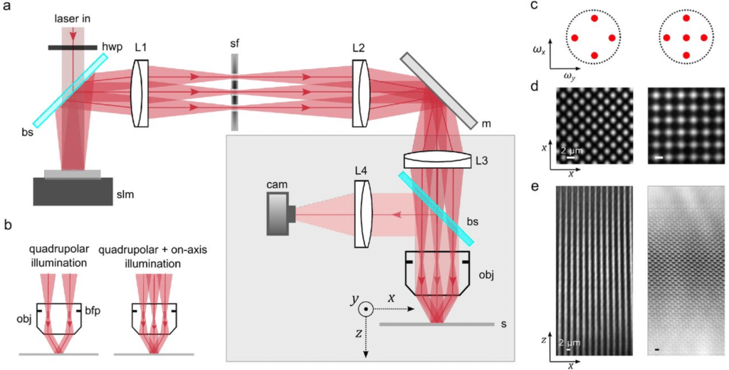
Polarization-engineered aberration-resilient light sheet microscopy
Yuqing Qiu, Juncheng Zhang, Christopher R. Warren, Sara Kacmoli, Vanessa Gonzalez, Cullen B. Young, Mengyang J. Li, Fei Liu, Kevin Keomanee-Dizon, Rebecca D. Burdine, Tian-Ming Fu
RAEM: random-access electron microscopy for revisitable 3D imaging
Ishaan Singh Chandok, Milan Patel, Yuelong Wu, Daniel Berger, Richard Schalek, Jeff W. Lichtman, Aravinthan D.T. Samuel, Yaron Meirovitch
High-resolution image-projection fluorescence lifetime imaging microscopy
WoongJae Baek, Jongchan Park, Liang Gao
Coherent Structured Illumination Microscopy with Enhanced Optical Sectioning
Kevin T. Crampton, Alan G. Joly, Long D. Nguyen, Saleem Iqbal, Robert W. Boyd, James E. Evans
Reflected Inline Detection in Epi Oblique Plane Microscopy
Md Nasful Huda Prince, Wishwa Herath, Balasubramanian Chellammal Muthubharathi, Nikhil Sain, Chitra Shaji, Md Rafiqul Islam Rupam, Aadil Qadir Bhat, Adil R. Wani, Mubarak Hussain Syed, Tae-Hyung Kim, Mark C. Walker, Olga Ponomarova, Tonmoy Chakraborty
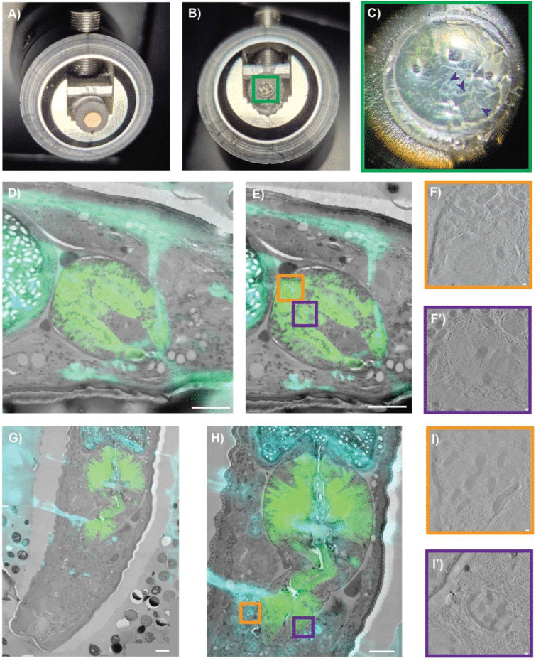
PinCorr: A high-pressure freezing carrier with intrinsic landmarks for cryo-correlative light and electron microscopy
Anna M. Steyer, Dietrich W. M. Walsh, Euan Pyle, Nadav Scher, Timo Zimmermann, Simone Mattei
Gold Electron Microscopy Grids with Anisotropic Foil Geometry Enable On-Grid Contact Guidance
Amit Avrahami, Noa Ben Asher, Ran Zalk, Leeya Engel
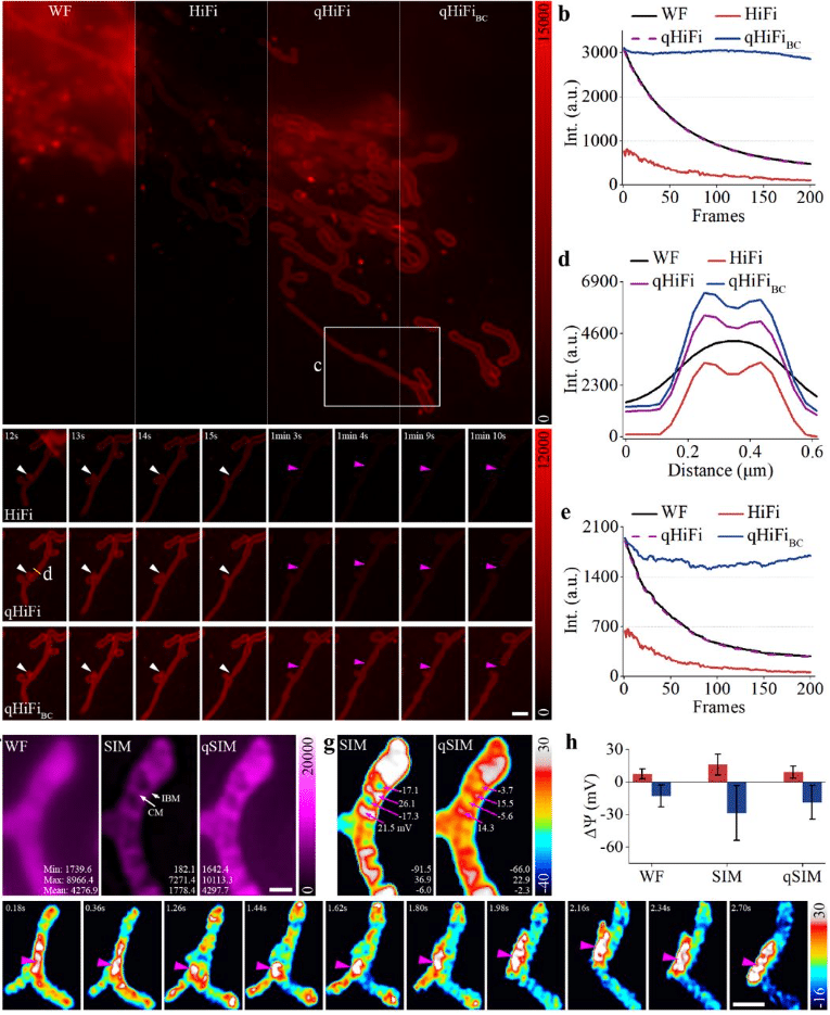
Physics-driven self-supervised learning for quantitative high-fidelity structured illumination microscopy
Yujun Tang, Zewei Luo, Xi Zhu, Wenyi Wang, Xichuan Ge, Meiqi Li, Cong Chen, Tongsheng Chen, Ceshi Chen, Peng Xi, Gang Wen
Triplet tumbling microscopy enables in situ quantification of protein complex assembly and dynamics
Julia R. Lazzari-Dean, Alfred Millett-Sikking, Prashant Rao, Zena D. Jensvold, Hannah Baddock, Maria Ingaramo, Aaron H. Nile, Andrew G. York, Magdalena Preciado López
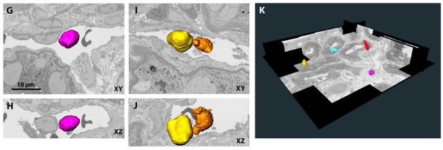
iSBEM: An Open-Source Workflow for Automated ROI Targeting in Volume Electron Microscopy
Paolo Ronchi, Graham Ross, Alana Burrell, Joost de Folter, Yanneck Klenz, Nedal Darif, Fiona Young, Matthew Lawson, Jonas Albers, Tobias Pietz, Friedrich Frischknecht, Elizabeth Duke, Candice Roufosse, Lucy Collinson, Amy Strange, Yannick Schwab
Evaluation of fluorescent proteins for compatibility with STED microscopy systems using two-color spectroscopies
Keisuke Sato, Daisuke Okada, Ayana Sugizaki, Tatsuo Nakagawa, Hiroshi Kumagai, Yoshinori Iketaki, Sumio Terada
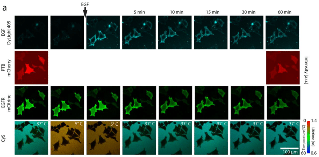
Robust thermometry-imaging at sub-micrometer and millisecond-resolution by fluorescence lifetime microscopy allows for additional acquisition of multiple imaging channels
Bijeesh Meethale Mangalassery, Simon Fabiunke, Malte Schmick, Jan Huebinger
HaloTag Ligand and HaloTag Protein engineering for a binary fluorescent turn-on probe
Blaise Gatin-Fraudet, Ulrich Pabst, Christina H. Olesen, Bianca C. Baciu, Ramona Birke, Sigrid Milles, Johannes Broichhagen
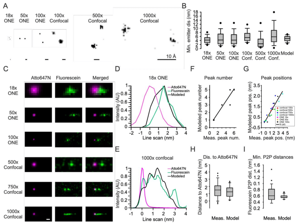
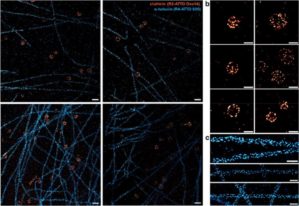
Two-dye-imager DNA-PAINT enables volumetric nanoscopy of expanded cells
Janna Eilts, Julia Weingart, Cerridwen Kiesel, Harsha Perozhy, Philip Kollmannsberger, Arindam Ghosh, Dominic A. Helmerich, Markus Sauer
Scalable Plasmonic Metasurface-Enabled Physics-Guided Self-Supervised Cellular Imaging
Cheng Zhang, susobhan choudhury, kerstin jansen, johannes balkenhol, Katrin Heinze
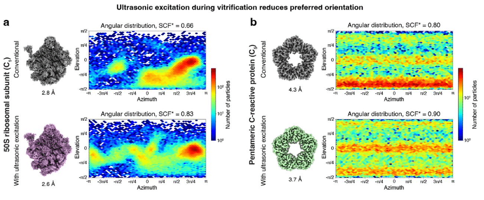
Overcoming Preferred Orientation in Cryo-EM With Ultrasonic Excitation During Vitrification
Harry M. Williams, Wyatt A. Curtis, Michal Haubner, Jakub Wenz, Marcel Drabbels, Ulrich J. Lorenz
High-speed volumetric single-molecule imaging using dual-wavelength light sheets and PSF-engineered enhanced biplane detection
Prakash Joshi, Nahima Saliba, Siyang Cheng, Yuya Nakatani, Dafei Xiao, Reut Orange-Kedem, Yoav Shechtman, Anna-Karin Gustavsson
Defocused reflectance imaging for low numerical aperture, large field of view quantitative live cell imaging studies
Sophie Bulloch, Tienan Xu, David Herrman, Paul Timpson, Tri Giang Phan, Yu-Hsuan Lin, Makoto Banno, Yean Jin Lim, Woei Ming Lee
Automated cryo-volume EM for high-resolution 3D imaging and in situ structural analysis of cells and tissues
Pavel Křepelka, Jana Moravcová, Zuzana Trebichalská, Elena Buglakova, Lenka Šmerdová, Hana Nedozrálová, Jaroslav Straník, Maria Rosario Fernandez-Fernandez, Pavel Plevka, Anna Kreshuk, Jiří Nováček
Volumetric nanoscale localization using engineered point spread functions in light sheet microscopy
R. E. Bautista Gonzalez, R. Mouthaan, A. Upadhya, D. J. X. Chow, K. R. Dunning, K. Dholakia
 (No Ratings Yet)