

Featured image with Manomay Pawar
Posted by FocalPlane, on 20 July 2026
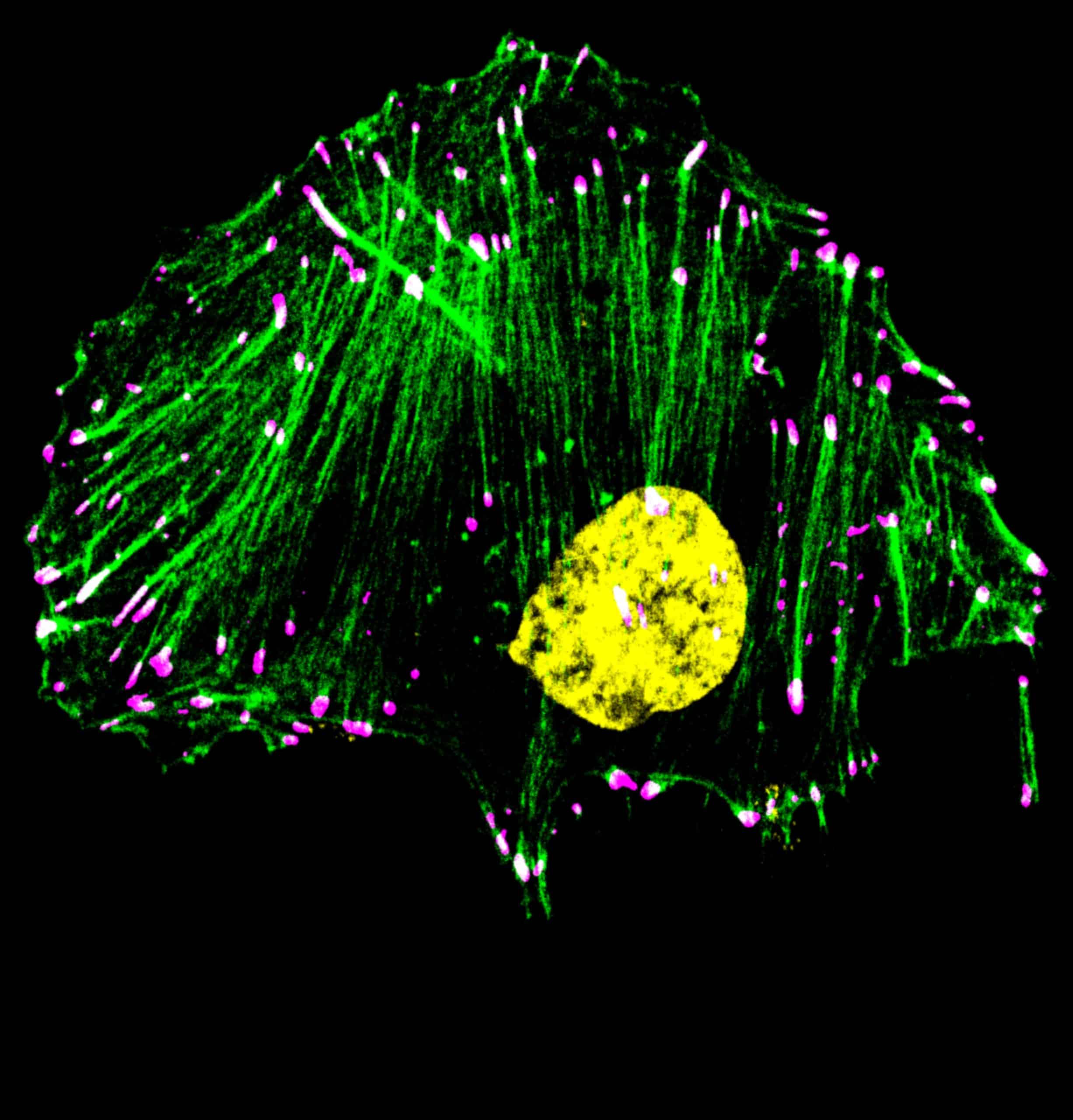
Our featured image, acquired by Manomay Pawar, depicts MDA-MB-231 triple negative breast cancer cells treated with drug Bemcentinib (R428), a small molecule inhibitor of AXL (a receptor tyrosine kinase) and labelled with phalloidin (actin;green), anti-Paxillin antibody to mark focal adhesions (magenta) and DAPI (DNA;yellow). The white coloured puncta represent colocalisation of paxillin with the ends of actin filaments. The sample was fixed with 3.5% PFA, followed by immunostaining. The image was captured using a ZEISS confocal microscope (Zeiss LSM 780) using a 63X oil immersion objective. It was processed/filtered using ImageJ software.
Discover more about Manomay’s research.
Research career so far: I am currently pursuing my Bachelor of Science-Master of Science (BS-MS) dual degree at the Indian Institute of Science Education and Research (IISER), Pune. My research journey began when I joined the Adhesion Lab, led by Dr. Nagaraj Balasubramanian, where I currently work as an undergraduate researcher. Looking ahead, I aspire to pursue a career in interdisciplinary oncology, focusing on the development of targeted cancer therapeutics. This scientific path, including the work featured here, is deeply personal to me. I dedicate this image and my research to my grandmother, who recently passed away from breast cancer. She is the profound driving force behind my work, and it is in her memory that I hope to make meaningful contributions to cancer biology in the future.
Current research: My current research focuses on understanding the role of the AXL receptor tyrosine kinase in regulating focal adhesion structure, specifically looking at its implications for cellular mechanosensing in human breast cancer cells. Because the mechanical environment dictates cancer progression, understanding this regulation could provide critical insights into how these cells develop metastatic capabilities, ultimately leading to novel therapeutic strategies. To investigate this regulation, I employ classic cell biology techniques alongside synthesizing 2D polyacrylamide hydrogels of physiologically relevant stiffnesses. This allows me to use advanced imaging to observe how breast cancer cells respond to their surrounding microenvironment.
Favourite imaging technique/microscope: Being in the early stages of my career, I have primarily used confocal microscopy for my studies, and I love its balance of simplicity and power. The feeling of watching a blurry frame suddenly resolve into an intricate cellular structure on the screen is truly surreal. Nevertheless, I am eager to expand my horizons by employing modalities like TIRF and SIM to further resolve the nanoscale architecture of focal adhesion complexes, allowing me to understand their structure even better.
What are you most excited about in microscopy? I am fascinated by our ability to visualize biological entities at the single-molecule level in live cells. I truly believe that seeing is the precursor to true understanding of any entity. Being able to track thousands of molecules simultaneously in a live cell would be a dream come true for any cell biologist. Beyond imaging hardware, I am also excited by the emergence of AI-driven plugins for image analysis and their ability to extract multi-parametric data from various kinds of samples. This synergy of high-resolution visualization and intelligent analysis is what excites me most about the future of microscopy.
Very nice Manomay