Technology highlights – Quantitative Phase Imaging (QPI)
Posted by Johanna Bischof, on 20 January 2021
Interview with Helena Chmelová, Ph.D. from the Light Microscopy Core facility at the Institute of Molecular Genetics of the Czech Academy of Sciences in Prague, Czech Republic.
Can you tell us a little bit about yourself – where you work, what your research focus is?
I work at the Light Microscopy Core Facility at the Institute of Molecular Genetics of the Czech Academy of Sciences in Prague as a light microscopy specialist. This facility is part of the Advanced Light and Electron Microscopy Node Prague of Euro-BioImaging.
I am particularly interested in imaging of living samples – cells, tissues and whole model organisms. One of my favourite imaging technologies is QPI microscopy. It’s an elegant method that is great for imaging living samples over extended periods of time with low phototoxicity and good cellular contrast.
QPI sounds fascinating. Can you please tell us about how QPI works and what it can be used for?
Quantitative Phase Image (QPI) microscopy is the spot-on method for long-term label-free imaging of living cells. As the light passes through a transparent specimen, different parts of the specimen with different thickness and distinct refractive indices generate slight changes in the phase of the light wave. In traditionally used methods, such as Phase Contrast or DIC, this phase shift is translated into the brightness variations in the image. These methods, however, do not extract any quantitative information out of it. QPI, on the other hand, provides quantitative information by creating a so-called phase image where acquired phase shift values are directly proportional to cell dry mass. Additionally, QPI produces artefact-free images (for example, it avoids the halo effect around the cells that is typical for Phase Contrast imaging) and therefore enables highly accurate segmentation of cells in the final image.
This method is very useful for studying cell shape, viability, motility, polarity, growth or differentiation under various conditions. A plethora of studies focusing on drug testing, cell interactions or cancer cell biology can take advantage of this technology.
Can you tell us a bit more about a specific project using QPI that you were involved with? What scientific questions were you addressing?
One of the projects which successfully exploited QPI technology in our facility focused on studying regulation of front-rear polarity in spreading cells. The authors wanted to evaluate reorganization of cell mass during cell polarization, therefore, QPI was the method of choice as it enables direct quantification of cell dry mass distribution and its changes over time. You can see an example image from their work in Figure 2 [1].
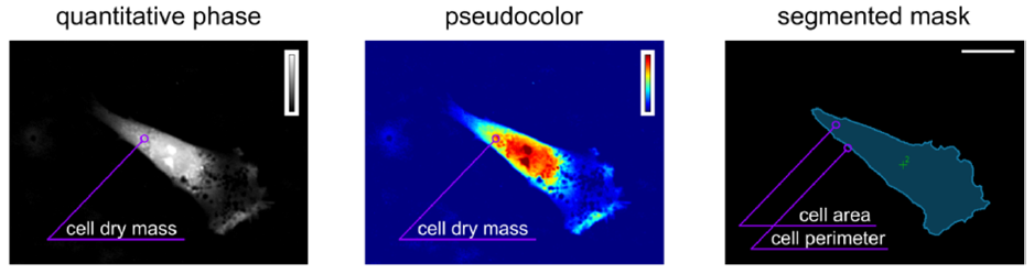
Image Credit: Tomas Grousl, Laboratory of Cell Signalling, Institute of Microbiology of the Czech Academy of Sciences.
Why was QPI the technology of choice here?
This technology enables visualization of low-contrast cells, offers direct observation of morphological changes, including rearrangement of cell mass, and facilitates subsequent image processing and analysis.
What are some challenges of using QPI? What do researchers have to pay attention to when performing these experiments?
This method is particularly convenient due to its low demands on sample preparation. As mentioned already, it enables users to image label-free cells without any need of contrast stains, which means that it is very gentle to the cells. Nevertheless, formation of the quantitative phase image can be influenced by the used media solutions and it is advisable to maintain stable conditions such as temperature, refractory index and constant volume of the solution while avoiding meniscus formation.
What other services do you provide in your facility that would be useful in combination with QPI microscopy?
We offer a wide range of light microscopy technologies from fluorescence widefield microscopy through confocal scanning or spinning disk microscopy to various super-resolution approaches. Hence, scientists here can build on their observation of cellular behaviour obtained by QPI imaging and easily dive into high-resolution techniques to further explore underlying mechanisms at subcellular or even single-molecule level.
Why should scientists visit your facility if they want to use QPI microscopy?
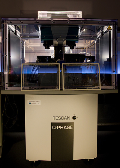
Our facility is equipped with the Telight Q-Phase (formerly TESCAN) coherence-controlled holographic microscope for quantitative phase imaging. Strikingly, we were privileged to be present at the development of this unique patented setup as the prototype of the machine was placed and operated in our facility in between 2014 and 2015. We therefore have a lot of experience in supporting users in their experiments with QPI microscopy. We are also in continued direct contact with the developers and we have their full support for our Q-Phase microscope and its users.
Importantly, Telight Q-Phase microscope provides QPI information directly by offering automated time-lapse segmentation and cell motility measurement in a very user-friendly software interface. Therefore, we believe that we can provide scientists with the comprehensive support ranging from the technical troubleshooting to the final analysis of acquired data. Additionally, Telight Q-Phase microscope enables us to combine QPI imaging with widefield fluorescence imaging which further broadens its versatility.
Want to use Quantitative Phase Imaging (QPI) via the Euro-BioImaging service?
Euro-BioImaging is the European landmark research infrastructure for biological and biomedical imaging as recognised by the European Strategy Forum on Research Infrastructures (ESFRI). All scientists, regardless of their affiliation, area of expertise or field of activity can benefit from Euro-BioImaging’s pan-European open access services. By facilitating user access to high quality imaging facilities, resources, and services, with a constantly evolving technology offer, Euro-BioImaging will boost the productivity and impact of research across Europe.
Potential users of the Euro-BioImaging technologies are encouraged to submit project proposals via our website. To do so, you can LogIn to access our application platform, choose the technology you want to use and the facility you wish to visit, then submit your proposal. By using Euro-BioImaging, users will benefit from advice and guidance by technical experts working at the Nodes, training opportunities, and data management services.
For more information visit our website at www.eurobioimaging.eu or contact us at info@eurobioimaging.eu

Helena Chmelová, PhD
Light Microscopy Core Facility
Institute of Molecular Genetics
Czech Academy of Sciences
Prague, Czech Republic
References
1. Ramaniuk, O., Klímová, Z., Groušl, T., Vomastek, T. (2020). Quantitative Phase Imaging of Spreading Fibroblasts Identifies the Role of Focal Adhesion Kinase in the Stabilization of the Cell Rear Biomolecules.10(8): 1089
 (No Ratings Yet)
Get involved
Create an account or log in to post your story on FocalPlane.
More posts like this


Filter by
- NewsApply
- DiscussionsApply
- How toApply
- ToolsApply
- Case studiesApply
- InterviewsApply
- JobsApply
- EducationApply
- Blog seriesApply
- WAMBIAN: West Africa.. in FocusApply
- Volume EMApply
- Latin American Micro..scopistsApply
- Bio-image Analysis w..ith NapariApply
- Imaging with…Apply
- Towards Global Acces..sApply
- Latin America Bioima..gingApply
- From Zero to Qupath ..HeroApply
- Asian Microscopists ..and Cell BiologistsApply
- AIC at HHMI JaneliaApply
- Deep Learning for Bi..o-image analysisApply
- GloBIAS – updates fr..om the communityApply
- Highlights from Euro..-BioImagingApply
- LSFM seriesApply
- DIY MicroscopyApply
- View all