Advertisement

Technology highlights - Traction Force Microscopy (TFM)

Posted by , on 9 December 2020

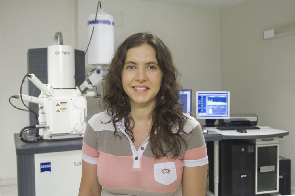
Interview with Aki Stubb, Ph.D. Please tell us a bit about yourself and the facility where you work. My name is Aki Stubb and I am a post-doctoral fellow at the University of Cambridge, UK. I did my PhD in the group of Johanna Ivaska at the Turku Bioscience Centre, University of Turku and Åbo

Technology highlights - Fluorescence Lifetime Imaging (FLIM)

Posted by , on 25 November 2020

Interview with Prof. Dolf Weijers and Prof. Joris Sprakel from Wageningen University, The Netherlands Tell us a little bit about who you are and about the people involved in the work we are talking about today. This work is a collaboration between the groups of Prof. Dolf Weijers and Prof. Joris Sprakel at Wageningen University,

Foldscope goes to the Peruvian Amazon!

Posted by , on 18 November 2020

Foldscope Instruments, Inc. is a company that was founded in 2016. We develop low-cost scientific tools with the goal of making science accessible to everyone around the world. In 2018, the Foldscope team visited Peru, Argentina, and Brazil. At the time, I was a Stanford graduate student in Biology and, due to school-related commitments, I

Technology highlights - Photo-acoustic Imaging (PAI)

Posted by , on 11 November 2020

Interview with Enzo Terreno, Ph.D. from the University of Torino, Italy. Please tell us a bit about yourself and the facility where you work. My name is Enzo Terreno, and I have the pleasure to be the Coordinator of the Multi-Modal Molecular Imaging (MMMI) Italian Node belonging to Euro-BioImaging. This is a multi-sited Node comprising

How to use Dragonfly Spinning Disk Microscope for Multiplex In Situ Hybridization

Sponsored by Andor, on 4 November 2020

Multiplexing in cell biology is the unveiling of several (Xn) RNAs in its 2D or 3D biological context. Multiplexing has become a hot topic in neurosciences, oncobiology, disease target diagnostics, development, behavioural studies, etc. Several techniques have been developed to allow multiplex imaging; examples of such are FISSEQ, instaSEQ, osmFISH, STARmap, MERFISH and seqFISH. The advantage of

Technology highlights - Coherent Anti-Stokes Raman Spectroscopy (CARS)

Posted by , on 28 October 2020

Interview with Antti Isomäki, PhD from the Biomedicum Imaging Unit of the University of Helsinki, Finland and Dalibor Pánek, PhD from the BIOCEV in Prague, Czech Republic. Tell us a bit about who you are and where your facility is based. AI: My name is Antti Isomäki and I am an optical physicist by training.

In-Situ Sequencing and Multiplex Imaging with the Dragonfly High Speed Confocal

Sponsored by Andor, on 27 October 2020

Challenge Background Understanding the molecular basis of development, brain function, neurodegenerative diseases, cancer, and behaviour is an enormous task. Up until recently, RNAs could be sequenced in bulk, or at the single-cell level, but unfortunately, the tissue environment information was lost. With smFISH [1] (single molecule FISH), the spatial information is retained, but this is

Microscopy Open Access collection 2020

Posted by , on 19 October 2020

Here on FocalPlane, Open Access Week is especially close to our hearts as it coincides with the birthday of the father of microscopy, Antonie van Leeuwenhoek (24 October). So every year for Open Access Week, we will be listing all the microscopy-related Open Access articles from The Company of Biologists journals on FocalPlane under the

Technology highlights - Spinning Disk Microscopy

Posted by , on 14 October 2020

Interview with Stoyno Stoynov, Ph.D. from the Center of Advanced Microscopy, Bulgarian Academy of Sciences, Bulgaria. Tell us a bit about the facility you run and what your focus is. The Bulgarian Node of Euro-Bioimaging ERIC is based in the Institute of Molecular Biology of the Bulgarian Academy of Sciences, Sofia. Our node is part

How I got into the Flamingo project, a new way of sharing custom-built light sheet microscopes

Posted by , on 12 October 2020

Sharing microscopy technology is crucial for biomedical research. This is the story how I joined Jan Huisken and his team to give other scientists quick access to custom-built light sheet microscopes.