/* */

Advertisement

An interview with Maria Eugenia Francia

Posted by , on 31 May 2022

Maria Eugenia Francia is a group leader at Institut Pasteur de Montevideo, focusing on Apicomplexan biology. Maria did her early studies (BSc and MSc) at the University of Idaho, where she started working on Apicomplexan biology. She then moved as a PhD student to the University of Georgia, where she continued this work. She later

Vote for your favourite image in the ELMI/FocalPlane microscopy image contest!

Posted by , on 30 May 2022

Voting for the image contest that we co-organised with the #ELMI2022 conference team is now open. We were thrilled to receive over 60 entries featuring a range of microscopy techniques, including widefield, confocal, spinning disk, super-resolution, and electron microscopy. VOTE HERE! Big thanks to all our contestants for their contributions and we wish them good

News digest

Posted by , on 27 May 2022

Here is a selection of interesting news, publications and discussions related to microscopy that happened in the last two weeks on Twitter and beyond. Talks, training and workshops: Abstract submission for the Royal Microscopical Society’s Abercrombie Meeting is open until 1 June. The meeting is in Oxford from 11-15 September 2022 and only takes place once

Explorative image data science with napari

Posted by , on 23 May 2022

When analysing microscopy image data of biological systems, a major bottleneck is to identify image-based features that describe the phenotype we observe. For example when characterising phenotypes of nuclei in 2D images, often questions come up such as “Shall we use circularity, solidity, extend, elongation, aspect radio, roundness or Feret’s diameter to describe the shape

Microscopy preprints – applications in cell biology

Posted by , on 20 May 2022

Here is a curated selection of preprints published recently. In this post, we focus specifically on preprints using microscopy tools in cell biology.

ELMI/FocalPlane image contest - deadline extended!

Posted by , on 20 May 2022

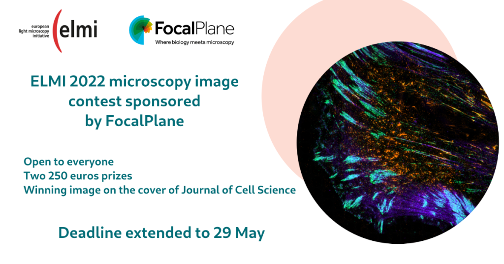
Does your work involve capturing stunning images on a microscope? Enter your image to our joint contest with ELMI (https://elmi2022.eu/) for a chance to win €250 and have your image featured on the cover of Journal of Cell Science. Competition details: The ELMI/FocalPlane image contest is open to anyone (not only participants of the ELMI

How Quantitative Phase Imaging can change the way you look at cells

Sponsored by telight, on 18 May 2022

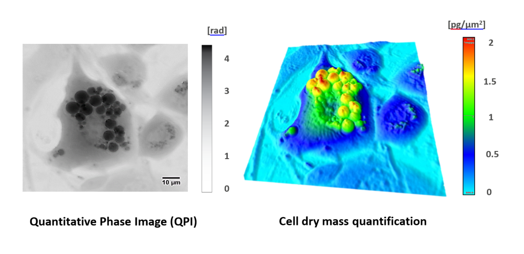
Looking at live cells is the key approach to cell analysis in both primary and applied research. But studying transparent living objects without disturbing them is challenging. Many of the commonly used methods for cell visualization are invasive and can affect results of an experiment. Even though these unwanted side-effects can be minimized due to

FocalPlane features... Quantitative Phase Imaging

Posted by , on 18 May 2022

Join us for our next FocalPlane Features… webinar on Thursday 30 June at 14:30 BST, sponsored by telight, to learn more about quantitative phase imaging and its applications to cell biology with Zuzana Nováková, Patrick McCall and Jan Balvan. The talks will be hosted in Zoom. Following the Q&A, there will be a round table

#GliaMorph - a tool to analyse glial cell morphology in 3D

Posted by , on 17 May 2022

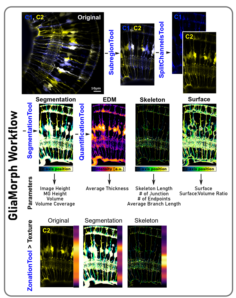
Contents What is #GliaMorph? What can you do and analyse with #GliaMorph? Links to manuscripts and data Contributions and Feedback Thanks to all contributors and people providing feedback References What is GliaMorph? GliaMorph is a toolkit implemented as Fiji Macros to allow modular application to process and analyse Müller glia morphology in the retina. During

An interview with Carolina Catta-Preta

Posted by , on 17 May 2022

Carolina Catta-Preta is currently a postdoc at Dr. David Sacks’ lab at the NIH (Bethesda, USA), where she is investigating Leishmania interactions with its insect vector, the sandfly. She started her career in Rio de Janeiro in 2007 at Carlos Chagas Filho Institute (UFRJ), in the Hertha Meyer lab under the supervision of Dr.