/* */

Advertisement

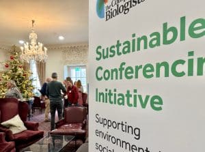
Support for your next sustainable scientific event in 2024

Posted by , on 9 January 2024

Scientific conferences and workshops play a crucial role in promoting collaborations and spreading awareness about new advancement done in the field. If you are interested in how to organise this type of events in a more sustainable manner, we are happy to support you.   If you do not know where to start, feel free

Featured images in 2023

Posted by , on 5 January 2024

In 2023, we started a featured image series and we have been delighted to highlight the microscopists that acquired these beautiful images! You can check out the full series here and in the gallery below. We are continuing the series in 2024 and would love to have nominations, including self-nominations. Send us an email at

Microscopy preprints – bioimage analysis tools

Posted by , on 29 December 2023

Here is a curated selection of preprints published recently. In this post, we focus specifically on new tools for bioimage analysis and data management released up until 20 December.

Crick Bioimage Analysis Symposium 2023 - a Review

Posted by , on 22 December 2023

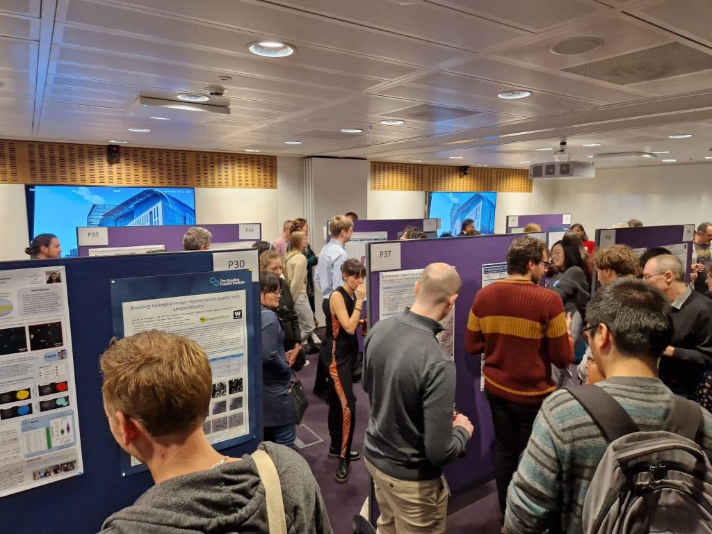
(By Vanessa Dao, Hradini Konthalapalli, Olatz Niembro Vivanco, Karishma Valand) The Crick Bioimage Analysis Symposium had its first in-person meeting in 2022. This year, #CBIAS2023 gathered around 200 people on site and 80 virtually. It has been an exciting two days of bringing biomedical researchers and their questions together with image analysis and their techniques.

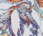
Featured image with Julien Rességuier

Posted by , on 22 December 2023

Our featured image, acquired by Julien Rességuier, shows the branchial cavity lymphoid architecture of an adult zebrafish. It is a 2D projection of a 3D multi-field of view acquisition of a transversal cryosection (30 μm) that has been stained with fluorescent phalloidin (inverted LUT – grey/copper) and DAPI (blue) to reveal tissue structures and labelled

StayGold variants explained!

Posted by , on 19 December 2023

I figured it might be useful as a reference, so here’s a blog-ified version of a thread I posted on X about recently published monomeric variants of the StayGold fluorescent protein. Thanks to Helen Zenner of FocalPlane for the help! In 2022, the Katayama/Miyawaki labs published StayGold, a bright and stable fluorescent protein (FP) from

Science Communications Officer at The Company of Biologists

Posted by , on 18 December 2023

We are currently recruiting for an exciting Science Communications role to enhance the community content on our journal websites and grow our social media presence in China through our WeChat channel.

Microscopy preprints – applications in biology

Posted by , on 15 December 2023

In this post, we share preprints that use microscopy tools to answer questions in biology.

Imaging with the Instituto Gulbenkian de Ciência’s Electron Microscopy Facility

Posted by , on 14 December 2023

In our ‘Imaging with…’ blog post, we meet the staff of the Electron Microscopy (EM) facility at the Instituto Gulbenkian de Ciência, Lisbon, Portugal.

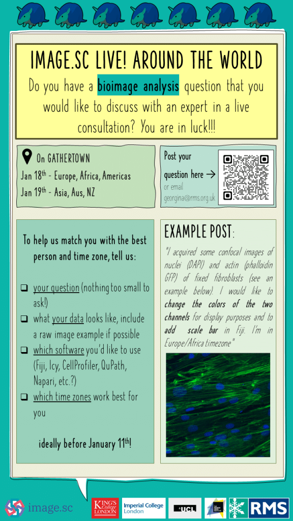
Image.sc LIVE! around the world - a live bioimage analysis helpdesk is coming to a time zone near you!

Posted by , on 13 December 2023

Community surveys often point to the biggest bottleneck in excellent bioimaging science being image analysis. That’s why the RMS DAIM committee and their friends across the world are putting the fantastic image.sc forum “on the road” and staging a first of its kind event which we’re calling “Image.sc LIVE! around the world”. All the info you need is here: Image.sc LIVE