Advertisement

Displaying posts with the tag: is_archive

AI4Life at the 5th NEUBIAS Conference

Posted by , on 26 May 2023

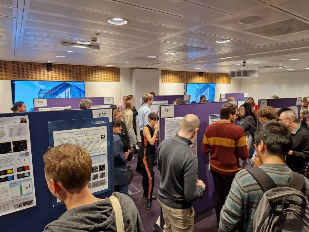
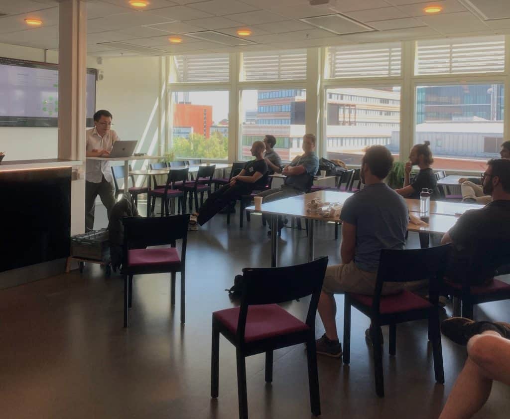
AI4Life recently participated in the 5th NEUBIAS Conference in Porto, Portugal, a two-part event comprising the Defragmentation Training School and the Open Symposium. This conference brought together experts and enthusiasts in the field of BioImage Analysis (BIA) to explore cutting-edge techniques and advancements in the integration of cloud computing. Under the umbrella of AI4Life, there

Feature extraction in napari

Posted by , on 3 May 2023

This blog post revolves around extracting and selecting features from segmented images. We will define the terms feature extraction and selection. Also, we will learn how to categorize features and can look up specific features in a glossary. Furthermore, we will explore how to extract features in napari. Definition of feature extraction During feature extraction,

Scientific Data Analysis: User Documentation 101

Posted by , on 30 April 2023

TL;DR: When publishing open-source tools for bio-image analysis special emphasis should be put on user-documentation. Users and developers have a different background and a language barrier limits knowledge exchange on how to use tools correctly. Writing a good user guide is a huge opportunity and worth the effort: Users get the most out of their

Quality assurance of segmentation results

Posted by , on 13 April 2023

This blog post revolves around determining and improving the quality of segmentation results. A common problem is that this step is often omitted and done rather by the appearance of the image segmentation than by actually quantifying it. This blogpost aims to show different ways to achieve this quantification as this leads to reproducibility. Therefore,

Annotating 3D images in napari

Posted by , on 30 March 2023

This blog post revolves around generating ground truth in 3D images for segmentation. Therefore, we will define what ground truth is and how we can generate it using napari in a time-efficient way. We will also learn about difficulties of annotating alone or in groups and address possible solutions for both. Challenges of image segmentation

Fast4DReg - to the rescue of your drifty microscopy data

Posted by , on 10 March 2023

Fast4DReg to the rescue of your 4D microscopy data In life sciences, researchers use microscopes to study living organisms, such as cells or small animals. These live cell imaging experiments are usually performed over several hours, exposing the experiment to changes in the sample and in the microscope surroundings – causing the data to drift.

Rescaling images and pixel (an)isotropy

Posted by , on 2 March 2023

This blog post shows the importance of rescaling 3D image data and what anisotropy of pixels/voxels has to do with it. We will see which different options exist to rescale an image as well as their advantages and limitations. We will also apply a commonly used segmentation algorithm, Voronoi-Otsu-Labeling, on the original and the rescaled

Microscopy preprints – bioimage analysis tools

Posted by , on 1 March 2023

Here is a curated selection of preprints published recently. In this post, we focus specifically on preprints on bioimage analysis, including an introduction to quantification in microscopy data from Siân Culley and colleagues.

Image Analyst at Harvard Medical School

Posted by , on 27 February 2023

We are looking for two or more smart, skilled, and enthusiastic bioimage analysts, to join us as soon as possible. Harvard Medical School (HMS) is a world leader in biological and biomedical imaging, with an outstanding community of researchers using cutting-edge microscopy to advance the field. The Image Analysis Collaboratory (IAC) is dedicated to training

AI4Life First Open Call

Posted by , on 20 February 2023

The European project AI4Life aims at narrowing the gap between life scientists performing biological imaging and developers of AI-based methods to analyze microscopy image data.  This is the first of a series of annual open calls, meant to provide life scientists who have unmet image analysis needs with adequate deep learning enhanced workflows for their