Featured image with Beste Senem Değirmenci
Posted by FocalPlane, on 18 August 2023
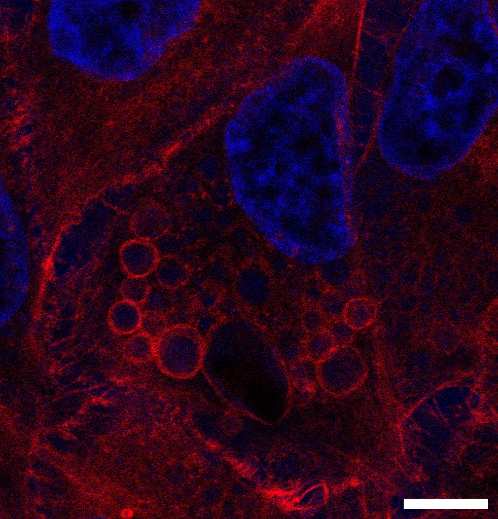
Our featured image, ‘Bubbles of The Cell’, shows enlarged endosomal vesicles in HeLa S3 cells upon inhibition of PI(3,5)P2 activity. PI(3,5)P2 is a crucial phosphoinositide involved in the regulation of endosomal trafficking. Depletion of PI(3,5)P2 disrupts the transition from early to late endosomes, leading to the enlargement of endosomal vesicles. Imaged with Leica DMI8 SP8 confocal microscope and processed with Fiji, ImageJ. Scale bar, 10 μm.
We caught up with Beste Senem Değirmenci to find out more about her research career and what she is excited about in microscopy.
Research career so far: I graduated from the Department of Molecular Biology and Genetics at Middle East Technical University (METU), Turkey in 2019. I was accepted to M.Sc. integrated Ph.D. program in the Department of Molecular Biology and Genetics at Koc University, Istanbul, Turkey in the same year. I joined Professor Nurhan Özlü’s lab, I am working on cell biology and proteomics for my research. I wish to continue my studies in the academic field as a post-doctoral researcher after my anticipated graduation in 2024.
Current research: I am investigating the molecular mechanism of the cell division-specific translocation of CLIC4 which has both soluble and membrane-bound forms in the cell. Our article showed that CLIC4 translocates to the mitotic cell surface and accumulates at the cleavage furrow during cell division (Uretmen Kagiali et al., 2020). My research aims to identify the translocation mechanism of CLIC4 during mitosis and cytokinesis.
Favorite imaging technique/microscope: I frequently use wide-field fluorescence microscopes and confocal microscopes for my research. I aim to improve the image quality in each experiment by testing various fixation methods and imaging settings. I attended several theoretical lectures about light sheet microscopes, super-resolution microscopes, electron microscopes, confocal microscopes, and fluorescence microscopes and I intend to go in-depth to understand more. Even though I couldn’t get experience with various microscopes, I am very curious about learning new imaging techniques in different microscope systems.
What are you most excited about in microscopy? I am fascinated by live-cell imaging which allows us to visualize the dynamic cellular process in real-time. The ability to capture proteins in action within living cells is inspiring to me. We can investigate the molecular mechanisms of the cell by imaging the fluorescence, which allows us to obtain protein localizations, the velocities of protein movements, and the lifetime of proteins. However, the time-lapse imaging of living cells is limited by spatial and temporal resolutions. Phototoxicity, image acquisition time, and optical resolution affect the image quality and maintaining healthy cells. Nevertheless, continuous advancements in imaging techniques, including high-resolution microscopes and improved image processing capabilities, enhance research quality by providing more detailed and accurate insights into cellular mechanisms, leading to a broader understanding of biological mysteries.
Uretmen Kagiali, Z. C., Saner, N., Akdag, M., Sanal, E., Degirmenci, B. S., Mollaoglu, G., & Ozlu, N. (2020). CLIC4 and CLIC1 bridge plasma membrane and cortical actin network for a successful cytokinesis. Life Science Alliance, 3(2), e201900558. https://doi.org/10.26508/lsa.201900558
 (1 votes, average: 1.00 out of 1)