Featured image with Elkhan Yusifov and Martina Schättin
Posted by FocalPlane, on 10 March 2023
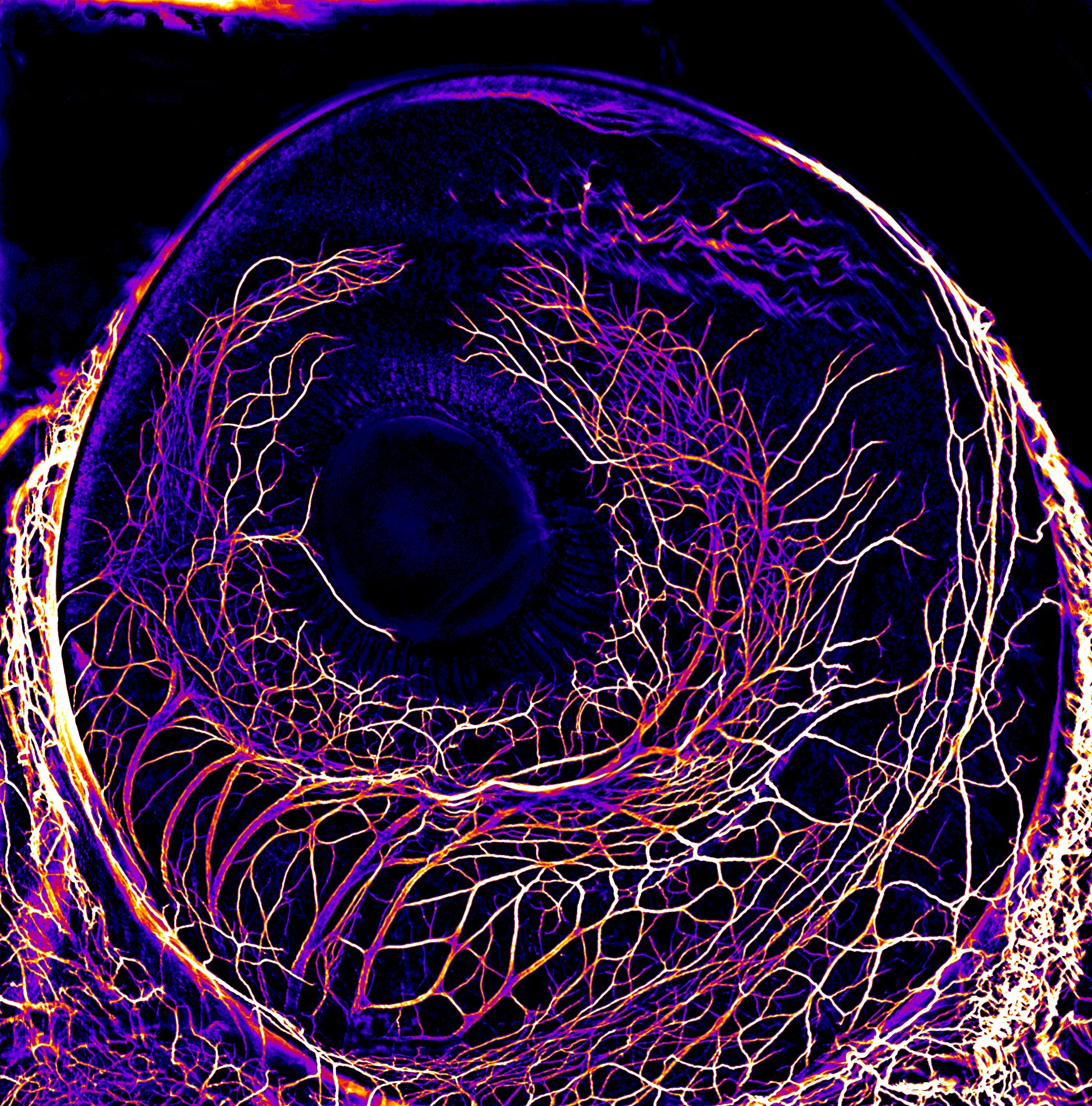
Our featured image, ‘Two eyes looking at an eye’, shows the developing neurons in the eye of a 7-day-old chick embryo, labelled with Neurofilament antibody using a whole-mount protocol (Voigt, et al. 2019). The embryo was cleared with BABB Solution and imaged on a Leica M205 FA microscope. The image was then post-processed with Fiji software and Fire LUT was applied.
We caught up with Elkhan Yusifov and Martina Schättin to find out more about their research and what they are excited about in microscopy.
Elkhan is a third-year PhD candidate in Professor Esther Stoeckli’s lab at the University of Zurich, while Martina completed her PhD in the Stoeckli lab in 2022 and is currently a postdoc in the lab of Professor Raghvendra Dubey at the University Hospital in Zurich (USZ). The Stoeckli lab, where the image was acquired, investigates axon guidance during neural development and studies the molecular mechanisms underlying the establishment of neuronal circuits.
Research career so far: I completed my bachelor’s degree with a double major in Neuroscience (I) and Cell and Molecular Biology (II) at the University of Toronto, Canada. I then moved to Zurich for my master’s degree in neuroscience where I am continuing with my PhD studies.
Current research: I am interested in the development of the nervous system. It is very exciting to see neurons and to work with them. Currently, I’m investigating how primary cilia, a tiny antenna in every post-mitotic vertebrate cell, contribute to nervous system formation. I am eager to learn both traditional and cutting-edge techniques to apply in my research.
Favourite microscopes: At the moment, my favourite microscopes are mesoSPIM and Talos TEM.
What are you excited about in microscopy? Microscopy allows us to discover the magnificent world of small structures at the highest resolution. Moreover, as a cherry on top of the cake for me, is that it is very exciting to be the first person ever to look at a thing and/or an event with microscopes.
Research career so far: As a master’s and then PhD student in the lab of Professor Stoeckli at the University of Zurich, I investigated the role of different axon guidance cues during neural circuit formation.
Current research: In the lab of Professor Dubey, we are dedicated to delineating the cellular and molecular mechanism(s) via which estrogen(s) induce angiogenesis and growth of cancer/tumour growth.
Favourite microscopes: Currently, I am amazed by the concept and image results of the MesoSPIM.
What are you excited about in microscopy? I enjoy microscopy a lot because it lets us dive into another world. New microscopy invention allows us to collect more knowledge and build up and/or rule out our hypothesis. I especially get a spark if, in one picture, you can not only clarify a scientific question but also be captured by the beauty of a single snapshot. At that moment, art and science come together in symbiosis.
 (No Ratings Yet)