Featured image with Upasana Gupta
Posted by FocalPlane, on 2 August 2024
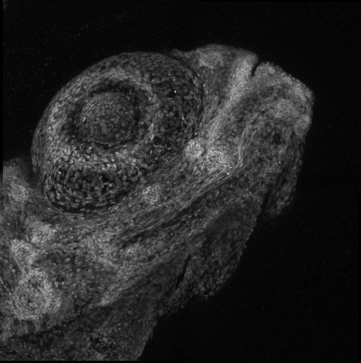
Our featured image, acquired by Upasana Gupta, represents the cellular architecture of zebrafish (larva) eye. The tissue was prepared by paraformaldehyde fixation of a 4-day post fertilisation larva, followed by staining with Hoechst nuclear stain. The sample was imaged and processed using Leica SP8 confocal microscope and Leica software at the Bio-imaging facility at the Division of Biological Science, Indian Institute of Science.

Find out more about Upasana’s research below:
Research career so far: I started out my research career during the final year of my undergrad. My first project at the Indian Institute of Science (Bangalore, India) was not a huge success but it was enough to hook me on to research was on how antibiotics can affect behaviour of an organism. The system I used was the classic genetics model, Drosophila melanogaster. Because I am a neuroenthusiast, I went on to continue my research in the same lab as a junior research fellow, researching how certain genes expressed in the neurons affect ageing. Four years into this project, I switched fields to biophysics where, in my PhD at the University of New South Wales (Sydney, Australia), I am addressing the kinetics of a pore forming protein during inflammation.
Current research: My current PhD project is on characterising the pore formation assembly kinetics of Gasdermin, which is an executioner of pyroptosis in mulitple cell types, including immune cells and neurons. In this project, we use a range of biophysical, biochemical, and cell biological approaches, including advanced fluorescence microscopy and single-molecule analysis. I am a little over a year in my PhD, waiting for some exciting results!
Favourite imaging technique/microscope: My favourite microscope so far is Andor Dragonfly Spinning Disc Confocal Microscope! Although I am using Total Internal Reflection Fluorescence Microscopy (TIRF-M) for my PhD project, most of my previous work involved long hours of confocal imaging.
What are you most excited about in microscopy? Life under a microscope means the world to me. The most exciting microscopy for me will be visualising real-time decision making in neurons! The brainbow of Zebrafish and Drosophila brain atlas projects are (for me) among the coolest neuronal imaging.


 (1 votes, average: 1.00 out of 1)
Excellent job and wish a great success ahead.