Microscopy preprints – applications in biology
Posted by FocalPlane, on 15 November 2024
Here is a curated selection of preprints published recently. In this post, we share preprints that use microscopy tools to answer questions in biology.
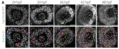
Timely neurogenesis enables increased nuclear packing order during neuronal lamination
Lucrezia C. Ferme, Allyson Q. Ryan, Robert Haase, Carl D. Modes, Caren Norden
All-Optical Multimodal Mapping of Single Cell Type-Specific Metabolic Activities via REDCAT
Yajuan Li, Zhaojun Zhang, Archibald Enninful, Jungmin Nam, Xiaoyu Qin, Jorge Villazon, Negin Fazard, Anthony A. Fung, Mina L. Xu, Hongje Jang, Nancy R. Zhang, Rong Fan, Zongming Ma, Lingyan Shi
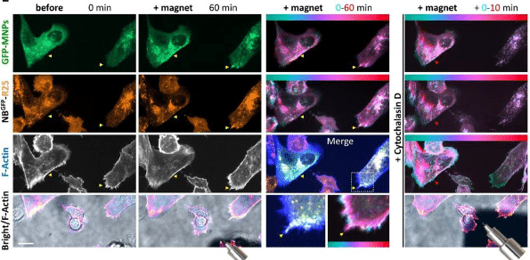
Live-cell magnetic micromanipulation of recycling endosomes reveals their direct effect on actin-based protrusions to promote invasive migration
Jakub Gemperle, Domenik Liße, Marie Kappen, Emilie Secret, Mathieu Coppey, Martin Gregor, Christine Menager, Jacob Piehler, Patrick Caswell
Complementation of a human disease phenotype in vitro by intercellular mRNA transfer
Gal Haimovich, Sandipan Dasgupta, Anand Govindan Ravi, Jeffrey E. Gerst
Triple labeling resolves a GPCR intermediate state by 3-color single molecule FRET
Leo Bonhomme, Ecenaz Bilgen, Caroline Clerté, Jean-Philippe Pin, Philippe Rondard, Emmanuel Margeat, Don C. Lamb, Robert B. Quast
Local optogenetic NMYII activation within the zebrafish neural rod results in long-range, asymmetric force propagation
Helena A Crellin, Chengxi Zhu, Guillermo Serrano-Nájera, Amelia Race, Kevin O’Holleran, Martin O Lenz, Clare E Buckley
Neurons and astrocytes have distinct organelle signatures and responses to stress
Shannon N. Rhoads, Weizhen Dong, Chih-Hsuan Hsu, Ngudiankama R. Mfulama, Joey V. Ragusa, Michael Ye, Andy Henrie, Maria Clara Zanellati, Graham H. Diering, Todd J. Cohen, Sarah Cohen
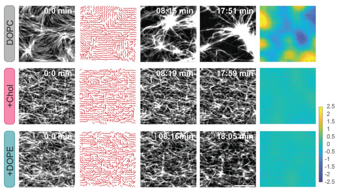
Anisotropic stretch biases the self-organization of actin fibers in multicellular Hydra aggregates
Anaïs Bailles, Giulia Serafini, Heino Andreas, Christoph Zechner, Carl Modes, Pavel Tomancak
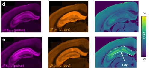
Brain-wide measurement of synaptic protein turnover reveals localized plasticity during learning
Boaz Mohar, Gabriela Michel, Yi-Zhi Wang, Veronica Hernandez, Jonathan B. Grimm, Jin-Yong Park, Ronak Patel, Morgan Clarke, Timothy A. Brown, Cornelius Bergmann, Kamil K. Gebis, Anika P. Wilen, Bian Liu, Ricard Johnson, Austin Graves, Tatjana Tchumatchenko, Jeffrey N. Savas, Eugenio F. Fornasiero, Richard L. Huganir, Paul W. Tillberg, Luke D. Lavis, Karel Svoboda, Nelson Spruston
Physical traits of supercompetitors in cell competition
Logan C. Carpenter, Shiladitya Banerjee
The A-C Linker controls centriole cohesion and duplication
Lorène Bournonville, Marine. H. Laporte, Susanne Borgers, Paul Guichard, Virginie Hamel
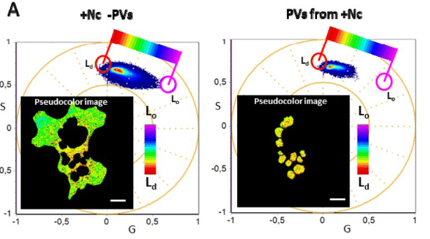
Modulation of Host Cell Membrane Biophysics Dynamics by Neospora caninum: A Study Using LAURDAN Fluorescence with Hyperspectral Imaging and Phasor Analysis
Marcela Díaz, Carlos Robello, Andrés Cabrera, Leonel Malacrida
Rho/Rok-dependent regulation of actomyosin contractility at tricellular junctions controls epithelial permeability in Drosophila
Thea Jacobs, Jone Isasti Sanchez, Steven Reger, Stefan Luschnig
Caging of membrane-to-cortex attachment proteins can trigger cellular symmetry breaking
Srishti Dar, Rubén Tesoro Moreno, Ivan Palaia, Anusha B. Gopalan, Zachary Gao Sun, Léanne Strauss, Richard R. Sprenger, Julio M. Belmonte, Sarah K. Foster, Michael Murrell, Christer S. Ejsing, Anđela Šarić, Maria Leptin, Alba Diz-Muñoz
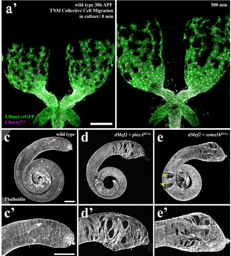
Plexin/Semaphorin Antagonism Orchestrates Collective Cell Migration, Gap Closure and Organ sculpting by Contact-Mesenchymalization
Maik C. Bischoff, Jenevieve E. Norton, Mark Peifer
Megakaryocytes assemble a three-dimensional cage of extracellular matrix that controls their maturation and anchoring to the vascular niche
Claire Masson, Cyril Scandola, Jean-Yves Rinckel, Fabienne Proamer, Emily Janus-Bell, Fareeha Batool, Naël Osmani, Jacky G. Goetz, Léa Mallo, Catherine Léon, Alicia Bornert, Renaud Poincloux, Olivier Destaing, Alma Mansson, Hong Qian, Maxime Lehmann, Anita Eckly
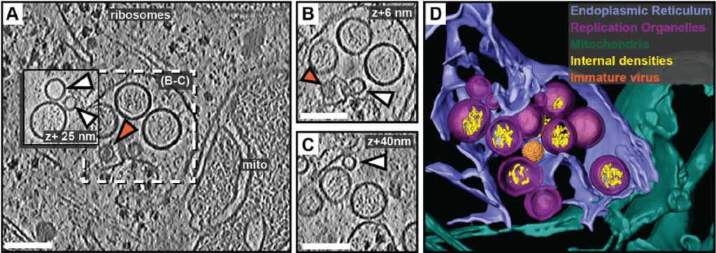
Cryo-electron tomography reveals coupled flavivirus replication, budding and maturation
Selma Dahmane, Erin Schexnaydre, Jianguo Zhang, Ebba Rosendal, Nunya Chotiwan, Bina Kumari Singh, Wai-Lok Yau, Richard Lundmark, Benjamin Barad, Danielle A. Grotjahn, Susanne Liese, Andreas Carlson, Anna K. Överby, Lars-Anders Carlson
Antagonism as a foraging strategy in microbial communities
Astrid K.M. Stubbusch, François J. Peaudecerf, Kang Soo Lee, Lucas Paoli, Julia Schwartzman, Roman Stocker, Marek Basler, Olga T. Schubert, Martin Ackermann, Cara Magnabosco, Glen G. D’Souza
Assessing Extracellular Vesicle Turnover In vivo Using Highly Sensitive Phosphatidylserine-Binding Reagents
Lavinia Flaskamp, Monica Prechtl, Annkathrin Scheck, Wenbo Hu, Christine Ried, Georg Kislinger, Mikael Simons, Anne Krug, Jan Kranich, Thomas Brocker
Origin of Ewing sarcoma by embryonic reprogramming of neural crest to mesoderm
Elena Vasileva, Claire Arata, Yongfeng Luo, Ruben Burgos, J. Gage Crump, James F. Amatruda
Visualizing nuclear pore complex plasticity with Pan-Expansion Microscopy
Kimberly J. Morgan, Emma Carley, Alyssa N. Coyne, Jeffrey D. Rothstein, C. Patrick Lusk, Megan C. King
Single Objective Light Sheet Microscopy allows high resolution in vivo brain imaging of Drosophila
Francisco J. Tassara, Mariano Barella, Lourdes Simó, M. Mailén Folgueira Serrao, Micaela Rodríguez-Caron, Juan Ignacio Ispizua, Mark H. Ellisman, Horacio O. de la Iglesia, M. Fernanda Ceriani, Julián Gargiulo
Holotomographic microscopy reveals label-free quantitative dynamics of endothelial cells during endothelialization
William D. Leineweber, Gabriela Acevedo Munares, Christian Leycam, Juliette Noyer, Patrick Jurney
Volume Electron Microscopy Reveals Unique Laminar Synaptic Characteristics in the Human Entorhinal Cortex
Sergio Plaza-Alonso, Nicolás Cano-Astorga, Javier DeFelipe, Lidia Alonso-Nanclares
Real-time high-resolution microscopy reveals how single-cell lysis shapes biofilm matrix morphogenesis
Georgia R. Squyres, Dianne K. Newman
Applying 3D correlative structured illumination microscopy and X-ray tomography to characterise herpes simplex virus-1 morphogenesis
Kamal L. Nahas, Viv Connor, Kaveesha J. Wijesinghe, Henry G. Barrow, Ian M. Dobbie, Maria Harkiolaki, Stephen C. Graham, Colin M. Crump
Ultrastructural expansion microscopy (U-ExM) visualization of malaria parasite dense granules using RESA as a representative marker protein
Junpei Fukumoto, Takafumi Tsuboi, Eizo Takashima
A combination of expansion microscopy and proximity labelling reveals conserved and unique asymmetric functional hubs at the trypanosome nuclear pore
Bernardo Papini Gabiatti, Johanna Odenwald, Silke Braune, Timothy Krüger, Martin Zoltner, Susanne Kramer
3D super-resolution imaging of PSD95 reveals an abundance of diffuse protein supercomplexes in the mouse brain
Sam Daly, Edita Bulovaite, Anoushka Handa, Katie Morris, Leila Muresan, Candace Adams, Takeshi Kaizuka, Alexandre Kitching, Alexander Spark, Gregory Chant, Kevin O’Holleran, Seth G. N. Grant, Mathew H. Horrocks, Steven F. Lee
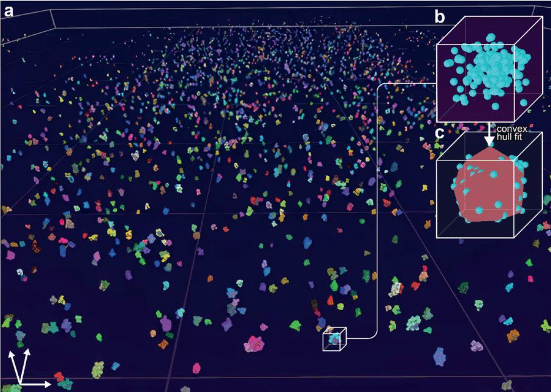
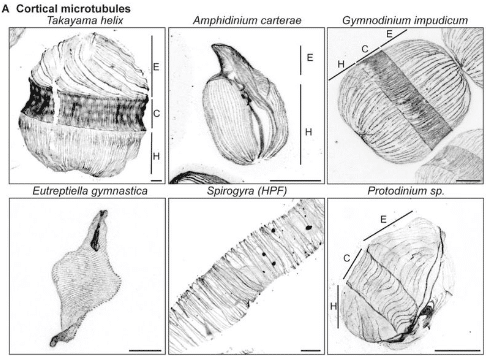
Charting the landscape of cytoskeletal diversity in microbial eukaryotes
Felix Mikus, Armando Rubio Ramos, Hiral Shah, Marine Olivetta, Susanne Borgers, Jonas Hellgoth, Clémence Saint-Donat, Margarida Araújo, Chandni Bhickta, Paulina Cherek, Jone Bilbao, Estibalitz Txurruka, Nikolaus Leisch, Yannick Schwab, Filip Husnik, Sergio Seoane, Ian Probert, Paul Guichard, Virginie Hamel, Gautam Dey, Omaya Dudin
Cryo-ET of actin cytoskeleton and membrane structure in lamellipodia formation using optogenetics
Hironori Inaba, Tsuyoshi Imasaki, Kazuhiro Aoyama, Shogo Yoshihara, Hiroko Takazaki, Takayuki Kato, Hidemasa Goto, Kaoru Mitsuoka, Ryo Nitta, Takao Nakata
Single fluorogen imaging reveals distinct environmental and structural features of biomolecular condensates
Tingting Wu, Matthew R. King, Yuanxin Qiu, Mina Farag, Rohit V. Pappu, Matthew D. Lew
Structuring Role of Tau-Tubulin Co-Condensates in Early Microtubule Organization
Chaelin Lee-Eom, Jaehun Jung, Celine Park, Min Ju Shon
Ultrastructural viscoelastic behavior of collagen identified by AFM nano-dynamic mechanical analysis
Meisam Asgari, Elahe Mirzarazi, Hojatollah Vali, Robert D. Frisina, Horacio D. Espinosa
Unwrapping the ciliary coat: high-resolution structure and function of the ciliary glycocalyx
Lara M. Hoepfner, Adrian P. Nievergelt, Fabrizio Matrino, Martin Scholz, Helen E. Foster, Jonathan Rodenfels, Alexander von Appen, Michael Hippler, Gaia Pigino
SMC modulates ParB engagement in segregation complexes in Streptomyces
Katarzyna Pawlikiewicz, Agnieszka Strzałka, Michał Majkowski, Julia Duława-Kobeluszczyk, Marcin Szafran, Dagmara Jakimowicz
Identification of nuclear pore proteins at plasmodesmata
T. Moritz Schladt, Manuel Miras, Jona Obinna Ejike, Mathieu Pottier, Lin Xi, Andrea Restrepo-Escobar, Masayoshi Nakamura, Niklas Pütz, Sebastian Hänsch, Chen Gao, Julia Engelhorn, Marcel Dickmanns, Gwendolyn V. Davis, Ahan Dalal, Sven Gombos, Ronja Lange, Rüdiger Simon, Waltraud X. Schulze, Wolf B. Frommer
A distinctive PI(4,5)P2 compartment forms during entosis and related engulfment processes
Joanne Durgan, Katherine Sloan, Marie-Charlotte Domart, Lucy M Collinson, Oliver Florey
Microtubule dynamics are defined by conformations and stability of clustered protofilaments
Maksim Kalutskii, Helmut Grubmüller, Vladimir A. Volkov, Maxim Igaev
Apical clathrin-coated endocytic pits control the growth and size of epithelial microvilli
Olivia L. Perkins, Alexandra G. Mulligan, Evan S. Krystofiak, K. Elkie Peebles, Leslie M. Meenderink, Bryan A. Millis, Matthew J. Tyska
Leukocytes use endothelial membrane tunnels to extravasate the vasculature
Werner J. van der Meer, Abraham C.I. van Steen, Eike Mahlandt, Loïc Rolas, Haitao Wang, Janine J.G. Arts, Lanette Kempers, Max L.B. Grönloh, Rianne M. Schoon, Amber Driessen, Jos van Rijssel, Ingeborg Klaassen, Reinier O. Schlingemann, Yosif Manavski, Mark Hoogenboezem, Reinier A. Boon, Satya Khuon, Eric Wait, John Heddleston, Teng-Leong Chew, Martijn A. Nolte, Sussan Nourshargh, Joachim Goedhart, Jaap D. van Buul
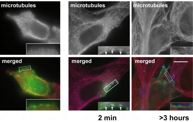
Mechanisms of cortical microtubule organization in epidermal keratinocytes
Keying Guo, Andreas Merdes


 (No Ratings Yet)