Featured image with Amy Engevik
Posted by FocalPlane, on 14 February 2025
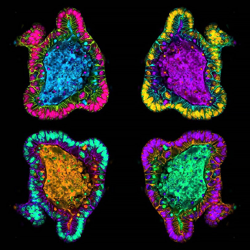
Our featured image, acquired by Amy Engevik, shows a differentiated intestinal organoid from a germline Myosin 5b knockout mouse.
How was the image prepared, imaged and processed? I generated intestinal organoids from small intestinal tissue, embedded the crypts in Matrigel and grew the stem cells to form ‘mini guts’ called organoids or enteroids. These organoids were fixed and paraffin embedded and then sectioned. I performed immunostaining to visualize the lateral membrane using a p120 antibody and a DPPIV antibody to show the apical and luminal region. Myosin 5b knockout mice exhibit striking intracellular inclusions that can be seen with DPPIV staining, and these organoids also show inclusions that are positive for DPPIV. I used photoshop to assemble pseudo-colored images and duplicated the image to vary the color scheme.
Discover more about Amy’s research:
Research career so far: I am currently a tenure track Assistant Professor at the Medical University of South Carolina in the department of Regenerative Medicine and Cell Biology. I study various aspects of gastrointestinal physiology and cell biology I did my PhD work at the University of Cincinnati studying gastric wound repair. My postdoctoral fellowship was at Vanderbilt University Medical Center. At Vanderbilt, I studied the pathophysiology of Microvillus Inclusion Disease, which results from mutations in trafficking proteins including Myosin 5b.
Current research: I study the epithelium of the gastrointestinal tract. One aspect of my lab is investigating how a high fat diet impacts epithelial cells in the GI tract and the other arm of my lab focuses on the role of Myosin 5b in GI health and disease.
Favourite imaging techniques/microscope: My favorite microscope is the Zeiss Axiocam.
What are you most excited about in microscopy? I’m excited about the use of nanobodies for immunostaining and about the single cell data sets that are becoming publicly available. I constantly use single cell data to check my staining accuracy to determine whether new antibodies are accurately labelling the correct proteins.
 (No Ratings Yet)