Featured image with Rohit Nautiyal
Posted by FocalPlane, on 9 May 2025
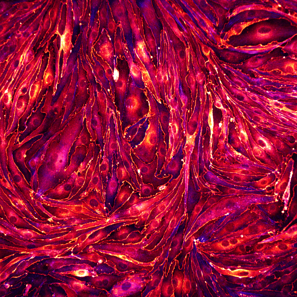
Our featured image, acquired by Rohit Nautiyal, shows a monolayer of brain endothelial cells seeded on a glass substrate coated with fibronectin. The cells were fixed and stained for ZO-1 (LUT- Red Hot) to visualise the tight junctions and actin (LUT-blue). The image was captured with a spinning disk confocal microscope and post-processed with Fiji software.
Discover more about Rohit’s research
Research career so far: My research journey began as a summer intern during my undergraduate studies at Lovely Professional University, Punjab where I was introduced to mammalian cell culture and cancer biology for the first time. Later, during my master’s degree at Birla Institute of Technology and Science Pilani, Hyderabad, I worked on a project focussed on optimising liposomal-mediated drug delivery in oral cancer cells. This exposure to a wide range of molecular and cellular techniques, motivated me to pursue a career in research. After completing my master’s, I transitioned from molecular biology to mechanobiology, an emerging field that bridges biology, engineering, chemistry, and physics. I joined Indian Institute of Technology, Goa as a Junior Research Fellow, where I worked on designing and fabricating micro-mechanisms for biaxial stretching of biological cells. Following this, I started my pre-doctoral research at Institute for Bioengineering of Catalonia (IBEC, Barcelona) in the Integrative Cell and Tissue Dynamics laboratory, led by Professor Xavier Trepat.
Current research: From the earliest stages of development and during life, cells self-organise within tissues, by actively coordinating their behaviour, guided by both chemical and physical cues. The process of self-organisation during morphogenesis involves a series of fundamental processes driven by cell mechanics. In these processes, cells change shape, divide, and move with respect to each other generating collective mechanical flows and forces stemming from the interplay between cell-substrate and cell-cell active and passive interactions. A crucial physical cue, affecting tissues composed of elongated cells, is multicellular orientation patterns, so called nematic patterns, which not only establish anisotropic mechanical properties but also well-defined active patterns of migration and force. As a doctoral researcher at IBEC Barcelona, I am studying the role of cell orientation and alignment as a mechanical cue for shape regulation in tissues.
Favourite imaging technique/microscope: The Olympus FV 3000 (laser scanning) was the first high-resolution microscope that truly amazed me with its incredibly detailed imaging. I still remember placing a leaf section under the microscope and being blown away by the intricate structure of the plant cells and stomata. They looked so much more fascinating in comparison to what I’d seen under a standard bench-top light microscope. After that, I became even more curious and started collecting samples from all around me – pollen, flowers, and even snake-skin – to see what other fascinating details the microscope could reveal. Each new sample brought even more stunning surprises!
In my doctoral research, I’m using the Nikon Eclipse series spinning disk confocal microscope to examine and study the mechanical role of alignment in endothelia. This technique allows me to capture the intricate structure and dynamics of these cells with incredible clarity.
What are you most excited about in microscopy? I’m currently excited about multichannel imaging (three or more channels) because it allows me to visualise different aspects of cells and their crosstalk by tracking multiple proteins under specific conditions. Also, I am excited about live imaging techniques, where we can see the normal development of cells into tissues and gain insights into cellular processes in real-time. Looking ahead, I’m especially interested in the advancements in AI-powered microscopy, particularly in areas like automated image analysis, cell segmentation, parameter quantification, and volumetric analysis at both single-cell and tissue levels.
 (7 votes, average: 1.00 out of 1)