Workshop report: 18th LFD Workshop in Advanced Fluorescence Imaging and Dynamics
Posted by FocalPlane, on 15 May 2025
The Laboratory for Fluorescence Dynamics ran their annual Advanced Fluorescence Dynamics Workshop in October 2024. Three of the attendees, Teemly Contreras, Irene Carlon-Andres and Razeen Shaikh were supported by JCS-FocalPlane Training Grants. They shared their experiences at the workshop here:
Teemly Contreras
The Advanced Fluorescence Dynamics Workshop organized by Laboratory for Fluorescence Dynamics (LFD) at the University of California Irvine was an exceptional opportunity to delve into advanced concepts of fluorescence imaging techniques and instrumentation. The workshop provided a comprehensive blend of theoretical lectures and hands-on laboratory training, fostering a deep understanding of state-of-the-art methodologies in microscopy and their applications. Supported by the JCS–FocalPlane Training Grant, my participation in this event enhanced my technical skills and scientific perspective.
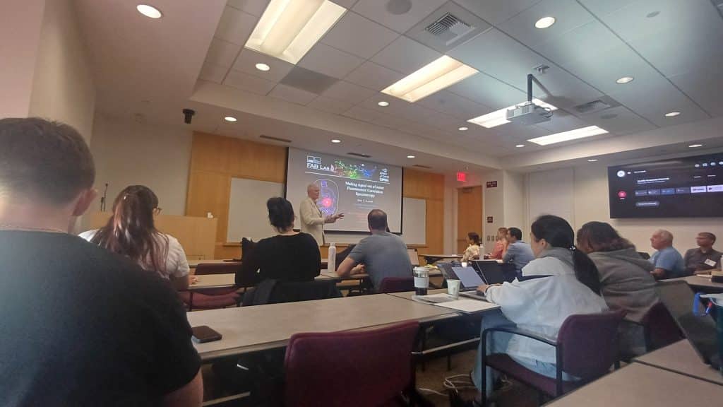
The workshop agenda was structured to maximize learning outcomes. The mornings were dedicated to theoretical lectures delivered by LFD scientists and distinguished invited speakers. Topics included Fluorescence Correlation Spectroscopy (FCS), Raster Image Correlation Spectroscopy (RICS), Fluorescence Lifetime Imaging (FLIM) and Förster Resonance Energy Transfer (FRET), Number and Brightness (N&B), Spectral Phasors and Super-Resolution Microscopy. The afternoons focused on practical sessions, where participants engaged with cutting-edge microscopy systems, including FLIM-Alba-STED, ONI Nanoimager, and FLIM Stellaris 8. These sessions were pivotal for gaining hands-on experience with advanced imaging techniques.

One of the most rewarding aspects of the workshop was the practical training. Participants were divided into small groups for an intimate and interactive learning environment. I was paired with a fellow researcher who studies T-cell Piezo channels, complementing my work on B-cell immune synapses. This collaboration facilitated insightful discussions on how various microscopy techniques could be leveraged to study immune synapse dynamics.
In addition to microscopy training, I attended a dedicated session on MESSR for image analysis. These sessions provided valuable insights into processing and interpreting fluorescence data, highlighting best practices for achieving reliable and reproducible results.
The workshop’s final day was uniquely structured, offering participants the freedom to interact with speakers and instructors. This open format allowed me to present my fluorescence images, receive constructive feedback, and discuss potential improvements. The positive feedback and guidance from experienced scientists were motivating and informed my ongoing research practices.
For me, this workshop was a dream realized. Many microscopy systems I encountered, such as FLIM and STED, were tools I had only read about in literature. Experiencing their functionality firsthand was both inspiring and enlightening. Moreover, the workshop underscored the potential of combining techniques, such as FLIM and STED, to achieve super-resolution imaging—a capability I plan to incorporate into my research on immune synapse formation. Beyond technical training, the workshop significantly expanded my professional network. Engaging with peers and experts in the field opened avenues for potential collaborations and enriched my understanding of global advancements in fluorescence microscopy.
I am deeply grateful to The Company of Biologists and the JCS–FocalPlane Training Grant for making my participation possible. This experience not only broadened my technical expertise but also reinforced my passion for microscopy and its applications in biological research. The knowledge and connections gained will undoubtedly have a lasting impact on my career, driving my contributions to the field of immunology and beyond.
In conclusion, the Advanced Fluorescence Dynamics Workshop provided an unparalleled platform to learn, innovate, and connect. I look forward to applying these insights to my research and sharing the knowledge with my scientific community.
Irene Carlon-Andres
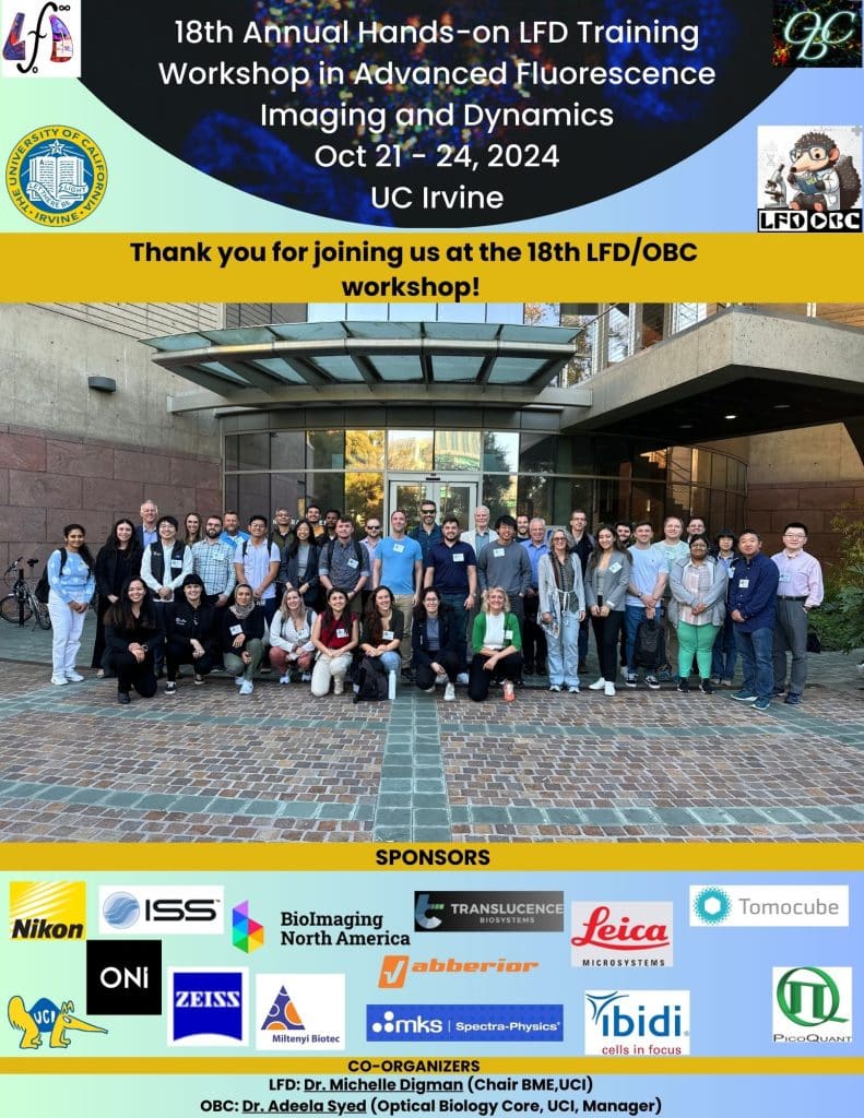
The Laboratory of Fluorescence Dynamics (LFD) in University of California Irvine (UCI) directed by Prof Michelle A Digman, together with the Optical Biology Core (UCI) managed by Dr. Adeela Syed, hosted the 18th Annual Hands-on training workshop in “Advance Florescence Imaging and Dynamics” held on the 21st-25th October 2024.
The workshop consisted of advanced theoretical lectures by LFD scientists and invited speakers, as well as laboratory and computer training. The topics included techniques based on fluorescence fluctuations, such as Fluorescence Correlation Spectroscopy (FCS); Fluorescence Lifetime Imaging (FLIM) and super-resolution microscopy. The theoretical lectures were conducted during morning sessions and introduced the principles of each technique with examples of applications in different areas of cell biology, infection and disease. The hands-on rotations were organized during afternoon sessions and focused on access to the microscopes located in the LFD and OBC facilities to learn about sample preparation and data acquisition, as well as training on open-access software specialized in fluorescence dynamics data analysis. The workshop was sponsored by renowned brands in the field of light microscopy who also provided demo systems, enabling the participants to have hands-on experience in cutting-edge technology.
At the end of each day, the participants had the opportunity to present their work to the audience in an informal setup, which facilitated the exchange of ideas about the potential and limitations of specific light microscopy techniques, data analysis methods and problem-solving driven discussions. I took the chance to present my current research, in which I aim to understand viral entry dynamics employing advance light microscopy techniques. In particular, I encouraged the discussion about possible routes for viral protein quantification using FCS. After my talk, I received very useful insight and feedback from the experts in the field.
The workshop had a well-balanced organization of the time employed for each activity. Free time was also scheduled between sessions, offering the opportunity to approach other participants and have one-to-one discussions. The number of attendees was limited to 50-100, which facilitated scientific exchanges. A social dinner event was organized during the second day at the University Club, offering also the possibility to visit the University campus.
The LFD organizes this workshop annually and it is a great opportunity to gain new skills and understanding in fluorescence imaging and analysis. It is also a great scientific environment to grow a network in the biophysics area, favouring the exchange and development of new interdisciplinary collaborations. I consider this experience has been enriching for my scientific career and I encourage other scientist to participate in future workshop sessions.
Razeen Shaikh
The LFD workshop was hosted by the LFD at UCI to disseminate advanced imaging techniques developed by them and other researchers in academia and industry around the world. The topics ranged from computational analysis of phasors to resolve imaging capabilities to development of advanced optics to visualize intracellular particles.
The workshop began with a tribute to Enrico Gratton who has contributed massively to the field of image processing.
On the first day, we were introduced to several techniques to quantify protein dynamics and estimate biophysical parameters in live tissues, including, diffusion rates and protein-protein interactions. These techniques include Fluorescence Correlation Spectroscopy (FCS), Raster Image Correlation Spectroscopy (RICS) and Number and Brightness (N&B).
On the following days, we were introduced to advanced microscopy techniques including fluorescence lifetime imaging (FLIM), stimulated emission depletion (STED), single molecule localization (SML), Hyperspectral imaging and image analysis techniques such as mean shift spatial resolution (MSSR).
These lectures were followed by hands-on rotation with, FLIM, two-photon microscopy, light-sheet and holotomography. There were several demonstrations by Zeiss, Leica and Nikon about cutting-edge advances made by their respective companies in advancing the ability to improve spatiotemporal resolution imaging, in the context of biology.
Additionally, we were introduced to methods such as expansion microscopy which employ chemical means to improve the ability to visualize intracellular bodies in cells.
All in all, the LFD workshop covered a diverse range of topics and initiated several stimulating discussions pertaining advanced imaging and analysis techniques.
I am grateful to the JCS–FocalPlane award for giving me this opportunity to attend the event.
Thank you!

JCS-FocalPlane Training Grants are open to all early-career scientists working in the field of cell biology. They can be used to attend training courses in microscopy and bioimage analysis. Drop us an email at focalplane@biologists.com if you have any questions about the grants.


 (No Ratings Yet)
Thank you alll so much for sharing and for your kind words! We’re thrilled to hear that you gained new skills and knowledge—and made new connections along the way. As Professor Enrico Gratton always said, “The true success of any workshop is measured by the curiosity and growth of its students.”
For those interested, the LFD Workshop has partnered with UC Berkeley’s AIM Workshop, now the Fluorescence Advanced Imaging Research (FLAIR) Workshop. We hosted it in Irvine this year, and next year it will be at UC Berkeley. We hope you’ll join us to explore advanced imaging technologies!